Alzheimer’s risk gene may boost memory in young

A fascinating study published in this month’s Cerebral Cortex reports that a gene known to massively increase the risk of Alzheimer’s disease in later life is associated, in young people, with better memory performance and more efficient use of the brain’s memory structures.

The research team, led by neuroscientist Christian Mondadori, looked at the genetics and memory performance of 340 volunteers, all in their early 20s.

The team were particularly interested in which version or allele of the apolipoprotein E (ApoE) gene each person had, because the ‘Epsilon 4’ allele raises the risk for Alzheimer’s disease in old age.

In fact, people with two ‘Epsilon 4’ alleles are virtually guaranteed to the brain disorder by the age of 80.

Each person took part in a word learning test that involved both short-term and long-term memory. This type of test is known to particularly rely on the function of the hippocampus, a key memory area which is known to decline in Alzheimer’s disease.

People who were carriers of the Epsilon 4 allele performed better in the long-term memory test, and no different for short-term memory.

The team decided to do more extensive memory tests while brain scanning 34 participants who were picked specifically to represent equal numbers of the three common genetic combinations.

These tests in the scanner involved learning faces and associations with professions over a number of trials and a target detection task that involved manipulating information in short-term memory (working memory).

There was no difference between the groups in terms of their accuracy on these tests, but people with the Epsilon 4 allele showed decreases in brain activity as time went on, suggesting they were using their brain more efficiently.

In contrast, people without the Epsilon 4 allele showed increases in brain activity, suggesting their brain was having to work harder to keep up.

A key question is why people who carry the Epsilon 4 allele would have a more efficient brain system for memory in early life but are more likely to have these same memory systems degrade in later life, as happens in Alzheimer’s disease.

As Alzheimer’s typically strikes after the time most people have children, the researchers suggest that the Epsilon 4 allele could confer an evolutionary advantage without adversely affecting chances of reproduction.

Some evidence that supports this idea has been found in previous studies where the ApoE Epsilon 4 allele has been associated with higher IQ scores, reduced heart activity under stress, and reduced chance of difficulties during pregnancy and post-birth problems.

Link to abstract of scientific study.

Old School Neurophysiology

squidaxon.jpgThe Plymouth Marine Laboratory brings us footage of experiments on the giant axons of the squid — the work that brought us the action potential. Quoting:

“The Squid and its Giant Nerve Fiber” was filmed in the 1970s at Plymouth Marine Laboratory in England. This is the laboratory where Hodgkin and Huxley conducted experiments on the squid giant axon in the 1940s. Their experiments unraveled the mechanism of the action potential, and led to a Nobel Prize. Long out of print, the film is an historically important record of the voltage-clamp technique as developed by Hodgkin and Huxley, as well as an interesting glimpse at how the experiments were done. QuickTime video excerpts from the film are presented here.

Link: excerpts from The Squid and its Giant Nerve Fiber

(via Three-Toed Sloth)

Discover 10 unsolved mysteries of the brain

Discover magazine has an article on ’10 unsolved mysteries of the brain’ which describes some of the biggest challenges in contemporary neuroscience.

It’s an interesting list, not least because you’ll notice that several of the problems are conceptual rather than empirical.

For example, the list includes ‘What are emotions?’, ‘What is intelligence?’ and ‘What is consciousness?’ that depend on a good philosophical analysis rather than just more data gathering.

In contrast, some of the other mysteries include things such as ‘How is information coded in neural activity?’ which is a problem of dealing with the complexity of the signals and their effect, rather than us having problems with defining any of the problem.

The fact that brain research relies as much on conceptual developments as laboratory work is one reason why philosophers are so important to cognitive science.

I like to think of them as conceptual engineers.

Link to Discover article ’10 Unsolved Mysteries Of The Brain’.

Brain scanning the dead

A study published in Forensic Science International has examined how brain scans can be of use to forensic pathologists – clinicians who perform autopsies to better understand how someone has died, often to provide evidence for a criminal investigation.

Head injuries are unfortunately common. Serious head injuries are most commonly caused by traffic accidents in Europe and Canada, while in the USA they are most commonly caused by firearms.

These cases will typically involve a police investigation, and the usual method is for a forensic pathologist to perform an autopsy on the head and brain to establish exactly what sort of injury occurred.

One of the drawbacks of this method is that it can only be performed once. The tissue is dissected and it’s not possible to keep anything except small samples.

This can be a problem in court, because it means the pathology evidence largely rests on a single examination, done in whatever way the pathologist thought was best, and can rely on their subjective interpretation.

A brain scan might be useful in this situation as it could be independently assessed and might actually pick up some things which might otherwise be missed if the head has to be dissected to be examined.

The study, led by Dr Kathrin Yen, compared findings from a structural MRI scan, a CT scan (an older structural brain scanning technique that uses X-rays) and a post-mortem, on 57 people, the majority of whom had died from serious head injury.

The findings from the scans and the autopsies were compared to see how well they agreed with each other.

The examination of the brain scans entirely missed some signs, such as increased brain pressure, but was 100% accurate in others, such as detecting bleeds between the dura mater, the brain’s tough outer membrane, and the skull.

The researchers note that some of the poor results are likely to be because radiologists aren’t used to forensic examinations as they’re trained to examine living people.

However, the brain scans had a distinct advantage in some cases. In one instance, the brain scan better estimated the size of an internal bleed which was exaggerated during autopsy because it bled further as the brain was cut.

The brain scans also allowed 3D reconstructions which could be examined from various angles to better understand how impacts occurred or what sequence of events might have caused the damage.

The image on the left is of a heat-induced fracture in a man who died in an aeroplane crash. The scan allows the pathologist to see the relationship between the skull fractures and the bleeds in the brain from a number of angles.

The study suggests that brain scanning corpses may give important clues in a forensic investigation but that radiologists may need to be specially trained so they know what they’re looking for.

Link to abstract of scientific study.
Link to more on forensic radiology from Radiology Today.

Brain electrodes ‘awake’ brain injured man

This week’s Nature has an intriguing short paper by a team of neuroscientists who ‘awoke’ a man from a ‘minimally conscious state’ by activating a surgically implanted brain electrode.

Like a coma, ‘minimally conscious state‘ (MCS) occurs after severe brain injury, but is not a state of complete unconsciousness.

Instead, the patient seems mostly unresponsive but can occasionally produce simple responses to commands or prompts that suggest inconsistent consciousness, such as short purposeful actions, brief verbalisations or emotional reactions.

Like coma, MCS is not a caused by a specific type a brain injury. It’s a description of the person’s behaviour, so it could be caused by varying damage to a wide range of brain systems.

However, it is known that MCS can be caused by problems with arousal. In other words, the major brain systems of the cortex might be relatively intact, but the system that regulates how active they are might be damaged, meaning the person has trouble staying conscious, despite having the potential for possible quite complex mental processes.

The frontal lobes, the brain stem and the thalamus are known to be key parts of the arousal system.

The man in question had been in a MCS for six years after suffering an assault with a blunt instrument that caused haematoma – bleeding in the brain, and subsequently, hydrocephalus – a build up of cerebral spinal fluid. Both of which put pressure on the brain that deformed and damaged it.

Because the man in question could intermittently respond to commands and give verbal responses, the researchers thought this might be a case where impaired arousal might be responsible.

To try and boost the activity in his arousal system, the team implanted a deep brain stimulation device (DBS) that sent electrical pulses directly into the thalamus via two brain electrodes.

After the initial tests, just 48 hours after surgery, there seemed to be some minor improvement in responses and EEG patterns.

This was a good sign, but because this was a new technique and each patient’s pattern of brain injury is distinct, the researchers had to then begin experimenting with different stimulation patterns.

After 18 weeks of testing, they found what seemed to be the optimal stimulation programme.

The patient showed longer periods of eye-opening and increased responsiveness to command and better limb control. He began to name simple objects, chew his own food and could produce up to six-word sentences.

In terms of the patient’s pre-injury level of functioning, the results are modest, but as an improvement on MCS, largely thought to be untreatable after 12 months, it’s a remarkable achievement.

The researchers note that this might not help all people with MCS, as this patient was specifically chosen because of his ‘widely preserved brain structure’, but, as with a previous treatment for coma we reported it’s more evidence that targeting the arousal system might be key in some cases.

In the same issue of Nature there’s an interesting (but closed access) commentary that makes some interesting points about what this tells us about consciousness, and particularly, the brain’s unconscious decision about when to rouse us into consciousness:

In essence, the brain does not process information in the abstract but instead consults information acquired through the senses and in memory insofar as it bears on the decisions made about potential actions and strategies. Our brains allow us to decide among possible options — that is, how and in what context to engage with the world around us. The brain makes many such decisions unconsciously. Indeed, the decision to engage at all is, in effect, an unconscious decision to be conscious. Thus, the brain of the sleeping mother queries the environment for the cry of her newborn. We suspect that the normal unconscious brain monitors the environment for cues that prompt it to decide whether to awaken and engage. This mechanism may be disrupted in various disorders of consciousness, including the minimally conscious state, hypersomnolence, concussion, abulia (lack of will) and possibly severe depression.

Previous theories of consciousness have relied on a central executive and magical physiological phenomena (for example, synchronized reverberations) to elevate the subconscious functions of the brain to consciousness. However, viewed as a decision to engage, consciousness can instead be studied in the same framework as other types of decision and the allocation of attention. Rather than a central executive, there seems to be a network of brain regions that organize the resting state and maintain overall orientation towards context. It is quite possible that they make decisions about whether or not to engage and in what way. They do what Sartre considered impossible: they choose whether to choose or not.

Link to Nature news story on the research (via Retrospectacle).
Link to summary of scientific paper.
Link to Nature commentary.
mp3 of Nature podcast discussion (starts 19 minutes in).
Link to write-up from ABC News.

An illustrated history of lobotomy

My last place of work blocked huge swathes of the web, meaning I’m discovering I’ve missed some blog posts recently, including this wonderful Neurophilosophy article on the rise and fall of prefrontal lobotomy.

It’s a fantastic tour through the history of how the procedure was developed, popularised and abandoned.

It aptly illustrates that medical history has been driven as much by personalities as by evidence, something which has only seriously been addressed in the last half-century by systematic trials and evidence reviews, largely due to the work of Archie Cochrane.

The article does have one quirk, where it equates early antipsychotic drug chlorpromazine with ‘psychosurgery gone wrong’.

Despite some serious and unpleasant side-effects (including movement disorders, sedation, weight-gain and dizziness), there is a large amount of evidence for its effectiveness in schizophrenia, and, in fact, was the first effective treatment for psychosis.

Even ignoring the brutal nature of the procedure, lobotomy was not even proven to be a useful ‘treatment’ by anything that would be accepted as reliable evidence today.

It is, however, an important chapter in the history of neuroscience, not least for what it tells us about how individuals can have such an influence on mainstream practice.

Link to article ‘The rise & fall of the prefrontal lobotomy’.

How gene therapy could cure brain diseases

Nature’s neurology journal has a freely available article on a technique that interferes with the translation of genetic information into proteins that may help prevent inherited brain diseases.

DNA has two main functions. The ‘template function’ of DNA is to pass on genes through generations and allow different traits to be inherited.

The ‘transcriptional function’ of DNA is to allow these genes to be expressed at appropriate times and places (and not expressed at others) to allow the cell to do its work.

‘Expression’ just means ‘turned into a protein’ and genes are just blueprints for proteins.

The blueprint gets turned into a protein by messenger RNA, which ‘reads off’ the information, then moves away to assemble the protein from a store of amino acid component parts.

As different cells in the body have different functions, and individual cells need to behave differently depending on what’s happening, different proteins need to be created at different times.

Disorders like Huntington’s disease result from genes that cause damaging proteins to be formed. These lead to the malfunction and death of brain areas that, in turn, leads to cognitive problems, movement difficulties, mental illness and eventual death.

Using a technique called RNA interference, researchers have found they can selectively interfere with the process where messenger RNA assembles proteins from the DNA’s genetic information.

Essentially, small chunks of gene-specific RNA are introduced into the cell, these find the messenger RNA and destroy the information before it gets turned into a protein.

In other words, it prevents specific genes from being turned into proteins.

This has caused a great deal of excitement because it could lead to treatments for disorders like Huntingdon’s by simply ‘silencing’ the rogue Huntingdon’s gene.

While you might have a rogue gene, RNA interference could essentially gag it, meaning it would never have a knock-on effect in the brain.

This has been demonstrated in very limited lab tests, and the Nature article examines the prospects for it being developed into a widespread treatment.

There are still some difficulties to overcome, however. One of which is how to get the interfering RNA into the right cells in the brain, a difficulty with many treatments owing to the filtering effect of the blood-brain barrier.

Another is how to make sure that the technique affects only the disease process. Researchers talk about proteins being involved in ‘chemical cascades’, meaning that they are involved in huge and complex mechanisms in the body.

It’s hard to predict exactly what effect silencing a gene will have, and whether your technique for doing so will also interfere with some other processes that use some of the same mechanisms, some of which we probably don’t even know about at the present.

RNA interference is still an experimental process, but it holds great potential for treating inherited brain diseases. The Nature article is a fantastic guide to the cutting edge of the science in this area.

Link to Nature Clinical Practice Neurology article on RNA interference.
Link to plain language guide to its use in Huntington’s.
Link to Wikipedia page on RNA interference.

Magnetic brain stimulation not proven to fight depression

At a recent American Psychiatric Association meeting, commercial companies were showing off custom made magnetic brain stimulators as a treatment for depression. A review article in the latest Nature Reviews Neuroscience looks at the technology and finds there’s still no convincing evidence that it’s an effective treatment.

The technology is based on transcranial magnetic stimulation (TMS), essentially a large electromagnetic which is activated near the scalp.

As you might remember from high school physics, a magnetic field that moves over a conductor causes a current. As your brain is a conductor, a current is formed in the neurons which cause them to briefly activate.

After an area of brain is magnetically activated, there are a few hundred milliseconds of inactive ‘silence’, effectively switching the area off, albeit safely and temporarily.

Depending on how quickly these pulses are applied, over a short period of time (typically a few minutes), the overall level of activity in the targeted brain area can be increased, or decreased. A technique known as repetitive or rTMS.

It has been known for a while that patients with depression have reduced activity in the left frontal lobe.

Researchers thought that TMS could be used to increase activity in this area and treat the depression, and so a long series of controlled trials were started to see how effective it could be.

It turns out, TMS does seem to reliably increase activation in the left frontal lobe, but the evidence on whether it actually improves depression in mixed, so mixed in fact, it’s not clear whether overall, it’s an effective treatment at all.

One of the difficulties is that there are so many variables to test out.

TMS can be applied to anywhere on the cortex, at varying strengths, at varying frequencies, at varying angles, with different wave forms and with different shaped coils, just to name a few of the possibilities that don’t include variation in the patients themselves.

Ridding and Rothwell, authors of the review paper, are not impressed with the results so far, but note some areas are promising but under-researched:

It is a sobering conclusion. A new treatment that might help some patients slightly more than placebo, but for which we do not know the most effective dose nor the best group of patients to target. Yet this is not the most worrying thing about the depression story. The main problem is that none of these trials has advanced our understanding of how rTMS may be having any action at all in depression. Trials currently underway are being conducted with almost the same rationale as the initial trials more than 10 years ago. The only changes are in variables such as the subset of patients being studied, or the intensity of the stimulus with respect to the distance of the patient’s brain from the scalp surface. In effect, the science has stood still.

In retrospect, depression was probably a poor choice of condition in which to begin trials of rTMS. It is phenotypically diverse with difficult diagnostic criteria and a subjective clinical evaluation that makes it highly susceptible to any placebo effects of rTMS. Diagnostically simpler conditions that have been studied more recently, such as auditory hallucinations in schizophrenia and tinnitus may prove more tractable. In both cases, rTMS of areas of the parietal or temporal cortices, respectively, have reduced symptoms, in some cases for several weeks after treatment. However, the number of studies done so far is small, and any firm conclusions about efficacy await much larger controlled trials.

This hasn’t stopped a number of companies producing ‘off-the-shelf’ TMS devices to make the technology more accessible to work-a-day psychiatrists, rather than clinical researchers.

There are currently some large scale trials being conducted to test further whether TMS for depression is a useful treatment, but so far, the evidence just isn’t there.

However, one promising avenue might be using TMS as a treatment for stroke – brain damage caused by bleeds and blockages in blood flow.

A different, but perhaps equally effective approach has been driven by a model in which recovery after stroke is suppressed in some patients by input from an ‘overactive’ non-stroke hemisphere. Reduction of the excitability of this hemisphere by low-frequency rTMS has also been reported to increase function, in this instance in a group of chronic patients whose stroke had occurred at least 1 year previously

It’s still early evidence, but it might be that using TMS to target specific symptoms and selective disorders may be more effective than trying to treat the diverse conditions that make up the common psychiatric diagnoses, such as depression, bipolar and schizophrenia.

Link to abstract of TMS review paper (sadly, not open-access).

Neuropsychopharmacology: The Fifth Generation

The American College of Neuropsychopharmacology have made a huge text book freely available online that covers the cutting edge of pretty much everything we know about how drugs affect the mind and brain.

Psychopharmacology is the science of how drugs affect the mind. You can do this without a huge understanding of brain function. You can just see how different drugs affect people’s mental state.

This was pretty much how many of the early drug treatments in psychiatry were discovered.

For example, the first antipsychotic, chlorpromazine, was developed in the 1950s as an antiemetic, a drug to prevent vomiting.

However, the French doctor Henri Laborit noticed that it induced a sort of ‘indifference’ to the world, and wondered whether it might help calm patients with mental illness who were agitated.

It was discovered that this drug was the first effective treatment for psychosis, and for several decades, psychopharmacology research simply tested various derivatives without a good understanding of how they were affecting the brain.

Neuropsychopharmacology adds neuroscience into the mix, and attempts to explain how drugs have their effect by studying how they interact with the biology of the brain.

It’s an incredibly important science, not only for the purpose of developing new treatments, but also for understanding how any drug (be it aspirin, cocaine or caffeine) has its effect.

The online text book, entitled Neuropsychopharmacology: The Fifth Generation reviews a huge, and I mean HUGE, amount of research into this area.

It’s an academic text, so is very in-depth, but is a fantastic resource to have freely available on the net.

Link to Neuropsychopharmacology: The Fifth Generation.

Virus linked to temporal lobe epilepsy

Open-access science journal PLoS Medicine published a recent study that suggests that infection with the herpes virus might cause temporal lobe epilepsy in some people.

The study found the virus in the brains of 11 out of 16 patients with temporal lobe epilepsy but not in those with other forms of epilepsy.

Studies that test brain tissue are often done post-mortem, on people who have died, because brain surgery is just too risky for the sake of removing samples for research.

This study is particularly impressive because it studied brain tissue from live patients.

In severe cases of epilepsy that don’t respond to medication, one option is to find which bit of the brain triggers the seizures (the ‘foci’) and surgically remove it.

This is particularly effective in people with mesial temporal lobe epilepsy, a type in which the foci is deep within the temporal lobes (mesial means ‘towards the midline’), usually stemming from disturbance in the hippocampus.

The team examined brain tissue removed in operations on 22 patients, and tested it for the presence of the human herpesvirus 6B (HHV-6B).

This type of herpes infection is incredibly common, more than 90% of the population have it. Normally, it’s completely harmless and just lies dormant in the body.

We don’t really know why, but in some people, it seems to reactivate, and is linked to neurological disorders like multiple sclerosis.

The researchers found that it was present in brain cells called astrocytes from 11 out of 16 patients with mesial temporal lobe epilepsy, but wasn’t present at all in patients with other types of epilepsy.

The image on the right is of a herpes infected astrocyte, the infection is visible due to a green marker.

They also studied one patient in more detail. He had four operations in a row, each of which reduced his seizures, until the final one left him seizure-free.

They found that the herpes virus was present most strongly in the temporal lobe tissue from the first operation, was weakly present in later operations, and wasn’t present in other brain areas.

They also found that infected brain tissue didn’t produce very much of a chemical that transports the key neurotransmitter glutamate across the brain.

If it doesn’t get transported properly, it ‘hangs around’, and because glutamate tends to make brain cells more active, too much could lead to overactivity and seizures.

To test the herpes – glutamate link, the team deliberately infected brain tissue taken from a patient without a previous infection.

In the lab, they discovered that herpes slowed the creation of the transporter chemical for glutamate, providing strong evidence for the link.

The evidence from the lab tests, the single case study, and the 22 patients, provides strong evidence that herpes infection could lead to temporal lobe epilepsy in some people.

This is an important finding because it suggests a cause for the disorder in some people, and provides a clear target which could lead to better treatments and prevention strategies.

What is still not clear is why this usually harmless infection might cause some people severe neurological problems, and remain dormant in others.

Link to PLoS Medicine paper.

Duped: Brain scan lie detection

There’s been quite a bit in the news recently about ‘brain scan lie detection’, but The New Yorker magazine have just published possibly the best article I’ve read so far on this intriguing but still-not-very-accurate technology.

It not only looks at the current technology, but also explores the dubious history of lie detection technology from times past.

The article is also remarkably well researched and level-headed, a balance that many stories about the technology sorely lack.

It points out some of the drawbacks of the technology, and some of frankly bizarre pitches being made by commercial companies.

One company recommends brain scans to help with “risk reduction in dating” and “trust issues in interpersonal relationships”!

Don’t get me wrong, people with brain scanners are sexy, but as with many things in life, it’s not what you have but what you do with it. Being shoved in a ‘fMRI lie detector’ by a potential lover would be a definite turn off.

The article is delightfully wide-ranging and talks to plenty of senior psychologists about their views on the technology and why we’re so attracted to brain scan evidence despite its drawbacks.

Really, an excellent piece. Well done New Yorker.

Link to New Yorker article ‘Duped: Can brain scans uncover lies?’.

Best of the Brain

A book called Best of the Brain from Scientific American (ISBN 1932594221) turned up unannounced the other day, and so far, I’m very impressed with it.

It’s a collection of twenty one of the most notable mind and brain articles from past issues of SciAm collected in a single volume.

I feel a bit reticent about waxing lyrical about a free book I’ve been sent, but I have to admit, I quite a fan of SciAm and SciAmMind, not least because they always make two feature articles from every issue freely available online which allows you judge the quality for yourself.

In fact, several of the articles from the book have already been made available online:

The Addicted Brain

Unleashing Creativity

Decoding Schizophrenia

Treating Depression: Pills or Talk

Controlling Robots with the Mind

Thinking Out Loud

Unfortunately, it doesn’t seem if the book’s a table of contents is online, as you could get a better idea of the diversity of topics that are covered.

Essentially, if you’re a fan of SciAm psychology and neuroscience writing, you’ll probably like this book. It’s really a greatest hits collection.

As this is the first unsolicited book I’ve been sent, a couple of clarifications. Readers: I’ll always say if a book I mention has been sent to me for free. Publishers: I won’t mention your book just because you’ve sent it to me.

Link to more info on the book.

The hardest cut: Penfield and the fight for his sister

In 1935, world renowned neurosurgeon Wilder Penfield published three remarkable case studies describing the psychological effects of frontal lobe surgery.

They remain a fascinating insight into the link between brain and behaviour, but one case was unlike anything Penfield had tackled before.

It described the fight to save the life of his only sister.


Continue reading “The hardest cut: Penfield and the fight for his sister”

Kidman new face of brain game, will it sharpen the mind?

As a sure sign that cognitive improvement games have gone mainstream, Nicole Kidman has been announced as the new face of Nintendo’s latest ‘brain training’ title.

The idea that mental training will actually help boost your mental skills is relatively new.

It was traditionally thought that the mind and brain just start losing their edge after young adulthood and your best hope was to learn to use your remaining resources more effectively as you age.

However, studies started to appear in the late 1990s suggesting that practicing certain tasks could act as a sort of ‘mental workout’, actually improving mental abilities directly in people with disorders like Alzheimer’s disease and schizophrenia.

Most people weren’t fully convinced of the benefits in healthy older people until a key study was published last year in the Journal of the American Medical Association that showed modest but reliable improvements, even after five years.

The effects were typically small (often too small to be picked up without standard tests), but interestingly, the training also had a knock-on effect on the participants’ ability to look after themselves effectively on a day-to-day basis.

It seems that cognitive training may have a stronger effect in people with mental impairments. A recent review of 17 studies found a positive effect on mental abilities, everyday activities and mood in people with Alzheimer’s.

However, as far as I know, no controlled trials have ever been published on any off-the-shelf ‘brain training’ game, including Nintendo’s. You’d guess from the medical literature that they might have a similar effect, but it’s yet to be shown for sure.

Link to BBC News article ‘Kidman to be new face of Nintendo’.
Link to JAMA article ‘Long-term Effects of Cognitive Training…’

Law, ethics, brain scans and mind reading

ABC Radio National’s All in the Mind has just
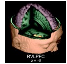
 broadcast the first of a two-part series on using neuroscience to read the mind.

The first programme investigates whether neuroscience can tell us anything about criminality and violence, and what role brain-based evidence will play in the court room.

The programme talks to many of the delegates from last April’s The Law and Ethics of Brain Scanning conference which was one of the first to consider the legal issues of brain scans in detail.

All of the conference talks have been put online as mp3 files so you can listen to the talks yourself if you want to hear more.

In the mean time, this edition of All in the Mind covers the key issues and next week’s will investigate some more (as yet undisclosed) aspects of so-called ‘mind-reading’ technology.

Link to AITM on ‘Mind Reading’.
Link to The Law and Ethics of Brain Scanning conference audio.

Labelling emotions reduces their impact

A brain scanning study has found that naming emotions reduces the intensity of emotion processing in the brain, possibly outlining a brain network responsible for the old saying ‘a problem shared is a problem halved’.

A team led by psychologist Dr Matthew Lieberman brain-scanned participants while they looked at pictures of faces that had different emotional expressions.

Earlier studies have found that naming an emotion seems to reduce its impact but this study went to particular lengths to make sure it was actually naming the emotion that helped, rather than just naming something, or identifying the emotion in other ways.

Participants were also scanned while having to name a face with a proper name, like Jane or Peter, or while matching the face to one with a similar emotional expression. This last task involved identifying the emotion but not naming it.

It turned out that when naming an emotion, and not for the other tasks, activity in a frontal lobe area called the the right ventrolateral prefrontal cortex (right VLPFC) significantly increased while activity in the amygdala decreased.

The amygdala is known to be heavily involved in processing emotions and seems to be regulated, at least in part, by the VLPFC.

These findings are consistent with this idea. The VLPFC increases its activity to dampen down the emotions triggered by the amygdala.

However, it’s not clear whether this happens equally for both positive and negative emotions, as 80% of the faces in the study had expressions of anger or fear, while only 20% displayed happiness or surprise, so this data only really tells us about unpleasant feelings.

We know that observing emotion in others makes us more likely to feel the same thing ourselves, but it’s not the same as experiencing an emotion ‘first-hand’, so we need to be a bit careful in assuming that this study fully represents the more everyday experience of talking about our emotions.

This experiment gives us a good understanding of the brain circuit involved reducing emotional impact via naming, but it doesn’t tell us much about why this occurs.

This is one of the major drawbacks of neuroimaging studies. They often just redescribe an effect in terms of brain activity.

Of course, this is essential knowledge, but we need to do more than just have several types of description and it is why the results from brain scanning studies need to be integrated with behavioural, experimental, clinical and subjective reports to be fully informative.

Link to write-up from APA Monitor.
Link to write-up from Scientific American.
Link to abstract of scientific study.