Detecting inner consciousness

CC Licensed photo by Flickr user hernán. Click for source.Mosaic has an excellent in-depth article on researchers who are trying to detect signs of consciousness in patients who have fallen into coma-like states.

The piece meshes the work of neuroscientists Adrian Owen, Nicholas Schiff and Steven Laureys who are independently looking at how to detect signs of consciousness in unresponsive brain-injured patients.

It’s an excellent piece and communicates the key difference between various states of poor response after brain injury that are crucial for making sense of the ‘consciousness in coma’ headlines.

One of the key concepts is the minimally conscious state which is where patients show signs of fleeting and impaired consciousness but which is nonetheless verifiably present.

However, MCS is still a very impaired state to be in and this is sometimes missed by news reports.

For example, lots of coverage of a recent Lancet study suggested that ‘one third of patients in persistent vegetative state (a state with no reliable signs of consciousness) may be conscious’ as if this meant they were fully conscious but trapped in their bodies, when actually they just reached criteria for minimally conscious state.

My only point of contention with the Mosaic article is that it’s a little too enthusiastic about sleeping pill zolpidem, which has been reported to lead to a ‘miraculous’ recovery in some case reports but where results from early systematic studies still look bleak.

Nevertheless, an excellent piece that’s probably one of the best accounts of this important and innovative area of research you’re likely to read for a long-time.
 

Link to Mosaic article ‘The Mind Readers’.

Coma alarm dreams

Intensive Care Medicine has published a wonderfully written and vivid account from a teenager who spent time brain injured and hallucinating in an intensive care unit.

The writer describes how he was admitted to intensive care at the age of 15 after suffering a head injury and had intense and bizarre hallucinations which are, as we know now, surprisingly common in critical care patients.

My experience of the time under sedation can be split into two. There was what I could perceive of the real world around me, and then there was my dream world.

In the real world, the most constant feature was sound. I could hear the nurses talking, understanding everything they said. They always spoke their names. They were always kind, conscious I think that I might hear them. They helped me to relax. I could hear the noises of the ward, tones of voices and alarms. The alarms made me tense. I can remember Mum talking to me a lot and Dad reading me ‘The Hobbit’, although I still can’t remember the names of all the dwarves. Mum and Dad’s voices always came from the left.

My other senses were not wholly switched off either. Things were put in my mouth: tubes, sucky things, wet watery pads and a toothbrush. Someone moved my hair about. I felt furry and silky toys placed under my fingers. My brother and sisters had brought a knitted tortoise and a horse for me. My feet were moved about and stretched, which felt really good. I remember that the rolled-up bed sheets were uncomfortable.

Other sensations were less good. The constant, repetitive shining of a bright light in my remaining eye really annoyed me – I am sure I can remember every single time.

Then there was my dreaming. I lived in the dream world nearly all the time and it went on and on. The dreams were vivid, terrifying and very disturbing. There were some good ones but unfortunately for me a lot of really bad ones. I can still remember most of them even now, more than a year since.

At the sound of an alarm, a giant monster appeared with a meat cleaver and pursued me around the sports hall. I had to protect a girl and prevent an army from crossing a river. The whole river and hall were aflame. I was burning from the heat.

In another I had to stop an alarm-driven colossal centipede from crossing a bridge. I could see the shadow of monsters looming towards me behind a curtain. I knew the monsters were there and about to consume me, but I lay transfixed, unable to move, and I remember feeling myself sweating with excruciating fear. I was then on the bridge of a nuclear submarine with maniacs trying to blow up the world, there was a huge explosion. Then it ended.

I was aboard a flying craft. I was there to stop green-coated aliens from creating human missiles. The aliens were forcing people into missile tubes. They were going to drop the human bombs from the aircraft.

Then there was a shape-shifter leopard beast chasing me and my friends. We were working in a fast-food place on a ship. It cornered us, and the Kentucky Fried Chicken sign burst into red lightning.

But I knew when something really nasty was going to happen. I could always hear the same alarm going off. It was a signal for the monsters to appear, for the centipede to attack, for bombs to be dropped, I would be sacrificed…I was very afraid. Tension would build to some hideous climax. Looking back, I suspect the pressure in my brain was causing both the nightmares and the alarm to go off.

I have made a great recovery from my injuries due in large part to the excellent care that was taken of my brain in intensive care. I have been into see the team a few times but I never stay too long. Those alarms still make me feel nervous!

As I noted in a recent article, these sorts of hallucinations were thought to be a distressing but ultimately irrelevant part of recovery but more recent studies suggests that have longer-term psychological impact that can be problematic in its own right.
 

Link to locked article ‘Coma alarm dreams on paediatric intensive care’

Bomb disposal for the brain

New Statesman has an excellent profile of the wise, funny and acerbic neurosurgeon Henry Marsh.

Marsh was the subject of the fantastic 2007 documentary The English Surgeon but he’s now one year away from retirement and has clearly decided that diplomatic responses are no longer a tactical necessity.

The piece also gives a vivid insight into the working life and daily challenges of a consultant neurosurgeon.

It’s also wonderfully written. This is pure joy:

When he finally went to medical school, at the Royal Free Hospital in London, he wasn’t sure about his choice. “I thought medicine was very boring,” he says bluntly. Henry is not a man to refrain from speaking his mind. “I didn’t like doctors. I didn’t like surgeons. It all seemed a bit dumb to me.” In Do No Harm he writes of his revulsion at what much surgery generally entails: “long bloody incisions and the handling of large and slippery body parts”.

But while working as a senior house officer, he observed a neurosurgeon use an operating microscope to clip off an aneurysm – a small, balloon-like blowout on the cerebral arteries that can cause catastrophic haemorrhages. It is intensely delicate work, using microscopic instruments to manipulate blood vessels just a few millimetres in diameter. It is also, as Henry says, like bomb disposal work, in that it can go very badly wrong – with the crucial difference that it is only the patient’s life at risk, not the surgeon’s. If this or any other kind of serious neurosurgery goes right, however, the doctor is a hero. “Neurosurgery,” he smiles, “appealed to my sense of glory and self-importance.”

Marsh has just written an autobiography called Do No Harm which I’ve just started reading. I’m only part way through but it’s already gripping and wonderfully indiscreet.
 

Link to New Statesman profile of Henry Marsh.

A balanced look at brain scanning

Bioethics think tank The Hastings Center have published an excellent open-access report on ‘Interpreting Neuroimages: The Technology and its Limits’ that takes a critical but balanced look at the use of brain scans for understanding the mind.

They’ve commissioned leading cognitive neuroscientists to write chapters including Geoffrey Aguirre, Martha Farah and Helen Mayberg, as well as having a chapter by some legal folks who discuss whether neuroimaging can teach us anything about moral and legal responsibility.

The chapter by the brilliant Martha Farah is particularly good and takes a level-headed look at the critiques of fMRI and is essential reading if you want to get up to speed on what brain scans are likely to tell us about the mind and brain.

The report is all in academic writing but if you’re a dedicated neuroscience fan, it probably won’t pose too much of a problem.


Link to ‘Interpreting Neuroimages: The Technology and its Limits’.

Frozen nightmares

The Devil in the Room is a fantastic short film about the experience of hallucinatory sleep paralysis – a common experience that has been widely mythologised around the world.

Sleep paralysis is the experience of being unable to move during the process of waking – when you have regained consciousness but you’re brain has not re-engaged your ability to control your muscles.

The reason the experience has been widely associated with mythological creatures is because in some people it can lead to intense emotions and hallucinations.

The name ‘sleep paralysis’ is a bit confusing because this also refers to normal sleep paralysis – where your brain disengages control of your muscles during REM sleep to stop you ‘acting out’ your dreams.

The film is part of the Sleep Paralysis Project, which has much more about the experience on their website.
 

Link to ‘Devil in the Room’ on vimeo.

Loving you is easy because you’re beautiful

Neuroscape Lab, we salute your next generation of brain visualisation, that looks like something out of a sci-fi film where the director is a bit obsessed with correctly representing the anatomy of the brain.

They describe the visualisation like this:

This is an anatomically-realistic 3D brain visualization depicting real-time source-localized activity (power and “effective” connectivity) from EEG (electroencephalographic) signals. Each color represents source power and connectivity in a different frequency band (theta, alpha, beta, gamma) and the golden lines are white matter anatomical fiber tracts. Estimated information transfer between brain regions is visualized as pulses of light flowing along the fiber tracts connecting the regions.

But honestly, who cares? It’s a glowing rotating brain with golden streaks of light flowing through it.

In fact, after 25 years, science has finally scanned the brain from The Orb’s ambient techno classic ‘A Huge Ever Growing Pulsating Brain That Rules from the Centre of the Ultraworld’.

It’s as if the rave generation stumbled out of life’s warehouse at 7am and ended up being neuroscientists.
 

Link to Neuroscape Lab’s awesome brain visualisation.
Link to the original Orb track (or the classic Orbital remix)

The Society of Mutual Autopsy

The Society of Mutual Autopsy was an organisation formed in the late 1800s to advance neuroscience by examining dead members’ brains and to promote atheism by breaking sacred taboos.

It included some of the great French intellectuals and radicals of the time and became remarkably fashionable – publishing the results in journals and showing plaster-casts of deceased members brains in world fairs.

In October 1876, twenty Parisian men joined together as the Society of Mutual Autopsy and pledged to dissect one another’s brains in the hopes of advancing science. The society acquired over a hundred members in its first few years, including many notable political figures of the left and far left. While its heyday was unquestionably the last two decades of the century, the society continued to attract members until the First World War. It continued its operations until just before World War II, effectuating many detailed encephalic autopsies, the results of which were periodically published in scientific journals.

The quote is from a fascinating but locked academic article by historian Jennifer Michael Hecht and notes that The Society was partly motivated by self-nominated ‘great minds’ who wanted to better understand how brain structure related to personal characteristics.

It was no backwater project and attracted significant thinkers and scientists. Most notably, Paul Broca dissected brains for the society and had his brain dissected by them, despite apparently never joining officially.

Part of the motivation for the society was that, at the time, most autopsies were carried out on poor people (often grave robbed) and criminals (often executed). The intellectual elite – not without a touch of snobbery – didn’t think this was a good basis on which to understand human nature.

Also, these bodies usually turned up at the dead of night, no questions asked, and no one knew much about the person or their personality.

In response to this, the Society of Mutual Autopsy functioned as a respectable source of body parts and also requested that members write an essay describing their life, character and preferences, so that it could all be related to the shape and size of their brain when autopsied by the other members.

There was also another motive: they were atheists in early secular France and they wanted to demonstrate that they could use their remains for science without consideration of religious dogma.

As with most revolutionary societies, it seems to have fallen apart for the usual reasons: petty disagreements.

One person took exception to a slightly less than flattering analysis of his father’s brain and character traits. Another starting flirting with religion, causing a leading member to storm off in a huff.

In a sense though, the society lives on. You can donate your body to science in many ways after death:

To medical schools to teach students. To forensic science labs to help improve body identification. To brain banks to help cure neurological disorders.

But it’s no longer a revolutionary act. Your dead body will no longer reshape society or fight religion like it did in 1870’s France. The politics are dead. But neither will you gradually fade away into dust and memories.

Jennifer Michael Hecht finishes her article with some insightful words about The Society of Mutual Autopsy which could still apply to modern body donation.

It’s “both mundane – offering eternity in the guise of a brief report and a collection of specimens – and wildly exotic – allowing the individual to climb up onto the altar of science and suggesting that this act might change the world”.
 

Link to locked and buried article on The Society of Mutual Autopsy.

A reality of dreams

The journal Sleep has an interesting study on how people with narcolepsy can experience sometimes striking confusions between what they’ve dreamed and what’s actually happened.

Narcolepsy is a disorder of the immune system where it inappropriately attacks parts of the brain involved in sleep regulation.

The result is that affected people are not able to properly regulate sleep cycles meaning they can fall asleep unexpectedly, sometimes multiple times, during the day.

One effect of this is that the boundary between dreaming and everyday life can become a little bit blurred and a new study by sleep psychologist Erin Wamsley aimed to see how often this occurs and what happens when it does.

Some of the reports of are quite spectacular:

One man, after dreaming that a young girl had drowned in a nearby lake, asked his wife to turn on the local news in full expectation that the event would be covered. Another patient experienced sexual dreams of being unfaithful to her husband. She believed this had actually happened and felt guilty about it until she chanced to meet the ‘lover’ from her dreams and realized they had not seen each other in years, and had not been romantically involved.

Several patients dreamed that their parents, children, or pets had died, believing that this was true (one patient even made a phone call about funeral arrangements) until shocked with evidence to the contrary, when the presumed deceased suddenly reappeared. Although not all examples were this dramatic, such extreme scenarios were not uncommon.

This sometimes happens in people without narcolepsy but the difference in how often it occurs is really quite striking: 83% of patients with narcolepsy reported they had confused dreams with reality, but this only happened in 15% of the healthy controls they interviewed.

In terms of how often it happened, 95% of narcolepsy patients said it happened at least once a month and two thirds said it happened once a week. For people without the disorder, only 5% reported it had happened more than once in their life.

Although a small study, it suggests that the lives of people with narcolepsy can be surprisingly interwoven with their dreams to the point where it can at times it can be difficult to distinguish which is which.

If you want to read the study in full, there’s a pdf at the link below.
 

Link to locked study at Sleep journal (via @Neuro_Skeptic)
pdf of full text.

Heroin, addiction and free will

The death of Phillip Seymour Hoffman has sparked some strong and seemingly contradictory responses. What these reactions show is that many people find it hard to think of addiction as being anything except either a choice or a loss of free will.

The fact that addiction could involve an active choice to take drugs but still be utterly irresistible seems difficult for most people to fathom.

Let’s take some reactions from the media. Over at Time, David Sheff wrote that “it wasn’t Hoffman’s fault that he relapsed. It was the fault of a disease”. On the other hand, at Deadspin, Tim Grierson wrote that the drug taking was “thoughtless and irresponsible, leaving behind three children and a partner”.

So does addiction trap people within its claws or do drug users die from their own actions? It’s worth noting that this is a politicised debate. Those who favour a focus on social factors prefer prefer the ‘trap’ idea, those who prefer to emphasise individual responsibility like the ‘your own actions’ approach.

Those who want to tread the middle ground or aim to be diplomatic suggest it’s ‘half and half’ – but actually it’s both at the same time, and these are not, as most people believe, contradictory explanations.

To start, it’s worth thinking about how heroin has its effect at all. Heroin is metabolised to morphine which then binds to opioid receptors in the brain. It seems to be the effects in the nucleus accumbens and limbic system which are associated with the pleasure and reward associated with the drug.

But in terms of motivating actions, it is a remarkably non-specific drug and it doesn’t directly cause specific behaviours.

In fact, there is no drug that makes you hassle people in Soho for a score. There’s no drug that manipulates the neural pathways to make you take the last 40 quid out of your account to buy a bag of gear. No chemical exists that compels your hands to prepare a needle and shoot up.

You are not forced to inject heroin by your brain or by the drug. You do not become an H-zombie or a mindless smack-taking robot. You remain in control of your actions.

But that does not mean that it’s a simple ‘choice’ to do something different, as if it was like choosing one brand of soft drink over another, or like deciding between going to the cinema or staying at home.

Addiction has a massive effect on people’s choices but not so much by altering the control of actions but by changing the value and consequences of those actions.

If that’s not clear, try thinking of it like this. You probably have full mechanical control over your speech: you can talk when you want and you can stay silent when you want. Most people would say you have free will to speak or to not speak.

But try not speaking for a month and see what the consequences are. Strained relationship? Lost job maybe? Friends who ditch you? You are free to choose your actions but you are not free to choose your outcomes.

For heroin addicts, the situation is similar. As well as the pleasurable effects of taking it, not taking heroin has strong, negative and painful effects.

This is usually thought of as the effects of physical withdrawal but these are not the whole story. These are certainly important, but withdrawing from junk is like suffering a bad case of flu. Hardly something that would prevent most people from saving their lives from falling apart.

For many addicts, the physical withdrawal is painful, but it’s the emotional effects of not taking drugs that are worse.

Most smack addicts have a frightening pre-drug history of trauma, anxiety and mood disorders. Drugs can be a way of coping with those emotional problems in the short-term.

Unfortunately, in the longer-term, persistent drug use maintains the conditions that keep the problems going. Even for those few that don’t have a difficult past or unstable emotions, life quickly become difficult after regular heroin use sets in.

If you can stay high, you’ll be less affected by the consequences of both long-standing problems and your chaotic lifestyle. If you stop, you feel the full massive force of that emotional distress.

It’s vicious circle that is often set in motion by past trauma but requires a meeting with a drug and the right social circumstances. Just taking the drug until you develop tolerance and withdrawal is unlikely to addict most people.

For example, a Vietnam War study found that just under half of soldiers reported trying heroin, 1 in 5 developed full blown dependency while in Vietnam but only about 5-10% of the dependent soldiers continued using when they arrived home. Most said they gave up without any help and only a small minority had ongoing addiction problems.

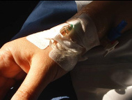
In fact, some of you reading this may have been addicted to heroin and not known it. Heroin, under its medical name diamorphine, is commonly used as a painkiller after major surgery. It’s not uncommon that patients develop tolerance and go into withdrawal after they leave hospital but just put it down to ‘feeling poorly’ or ‘recovering’.

But for persistent addicts, the ‘short-term solution that maintains the long-term problem’ cycle is not the whole story and it’s important to remember the neurological effects of the drug and how it interacts with, and changes, the brain.

Addiction is associated with difficulties in resisting cravings and making flexible decisions. This is likely to be caused by a combination of genetics, earlier experience and the ongoing impact of the drug and the drug-focused lifestyle – all of which affect brain function.

A recently popular approach is the ‘disease model’ of addiction which says that the brains of those who become addicted are more susceptible to compulsive drug use because of genetic susceptibility and / or brain changes due to early experience that ‘prime’ the brain for addiction.

It’s probably true to say that the extreme version of the ‘disease model’ – which says addiction is entirely explained by these changes and is best characterised as a ‘brain disease’ – is an exaggeration of what we know about the neuroscience of addiction, but this is not to say that neuroscience is not important.

But either way, there is no clear relationship between an aspect of behaviour being best explained in neurobiological terms and not having any control over that behaviour. For example, most genuine addicts usually give up, on their own, without any assistance and don’t relapse. They still have brains, of course.

Unfortunately though, the ‘disease model’ approach is often used precisely because some think it implies addicts have less control, possibly because they feel (probably wrongly) that it is less ‘stigmatising’ to think of heroin users in this way.

Instead, we know that self-efficacy is one of the best predictors of recovery, so denying people’s role in their own decisions just undermines one of their most important tools for recovery – alongside medication, social support and other forms of therapy.

So to say an addict has ‘no choice’ over their actions is just to misunderstand addiction but to pretend these choices are like any others just misses the fact that they can sometimes be impossibly hard decisions.

Unfortunately though, people find it hard to separate any admission of addicts being able to choose their actions from blame and moral accusation.

Blaming someone for their addiction is like shaming someone for being wounded by an abusive partner. Whatever the circumstances that caused the problem, they deserve respect and treatment, and working with them to help them regain control of their circumstances and promote their own autonomy is an important and valuable way forward.

The cutting edge of brain science technologies

National Geographic has an excellent article that gives a tour of some of the latest technologies of neuroscience that are likely to be leading the way in understanding the brain over the next decade.

You can read the full article online but you need to complete a free registration first. A typical publication ploy but, in this case, it’s well worth doing.


The article is itself fascinating but is also wonderfully illustrated with photos and videos to show exactly how the new technologies allows us to see the brain at work in many different ways.

An excellent guide to the cutting edge of lab brain science.
 

Link to National Geographic article ‘Secrets of the Brain’ (free reg required).
Link to plain text copy of article – no reg required.

The pull for lobotomy

The Psychologist has a fascinating article by historian Mical Raz on what patients and families thought about the effects of lobotomy.

Raz looks at the letters sent between arch-lobotomist Walter Freeman and the many families he affected through his use of the procedure.

Contrary to the image of the ‘evil surgeon who didn’t care about the harm he was doing’ many patients and families gave warm and favourable feedback on the effects of the operation.

Even some very worrying details about the post-operative results are recounted in glowing terms. Freeman had every reason to suspend his disbelief.

What it does illustrate is how a damaging and useless treatment could be perceived as helpful and compassionate by Freeman and, presumably, other doctors because of how docility and, in some cases, genuine reduced distress were valued above the person’s self-integrity and autonomy.

An interesting and challenging article.
 

Link to ‘Interpreting lobotomy – the patients’ stories’.

A multitude of phantoms

A fascinating paper in the neuroscience journal Brain looks at artistic depictions of phantom limbs – the feeling of the physical presence of a limb after it has been damaged or removed – and gives a wonderful insight how the brain perceives non-functioning or non-existent body parts.

In fact, most people who have a limb amputated will experience a phantom limb, although they often fade over time.

However, the feeling is usually not an exact representation of how the actual limb felt before it was removed, but can involve curious and sometimes painful ‘distortions’ in its perceived physical size, shape or location.

The Brain article looks at the diversity of phantom limb ‘shapes’ through their visual depictions.

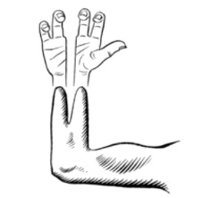
The image on the left is from a 1952 case report where an amputation involved a ‘Krukenberg procedure‘.

This operation is rarely performed in the modern world but it involves the surgeon splitting the stump to allow pincer movements – and in this case it left the patient with the feeling of divided phantom hand.

In other cases, without any out-of-the-ordinary surgical procedure, patients can be left with a phantom that feels like the middle parts of their limb are missing while they still experience sensations in phantom extremities.

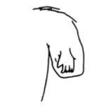
The drawing on the right was completed by a patient in a medical case study to illustrate their experience of a post-arm-amputation phantom limb.

In this case, the person experienced the feeling of a phantom hand on their shoulder stump, but had no experience of an intervening phantom arm.

While phantom limbs are usually associated with amputations, the phenomenon is actually caused by the mismatch between the lack of sensory input from the limb and the fact that the brain’s somatosensory map of the body is still intact and trying to generation sensations.

This means that any sensory disconnection, perhaps through nerve or spinal damage, can cause the experience of a phantom limb, even if the actual limbs are still there.

In the drawing on the left, a patient who suffered spinal damage that caused a loss of sensation in their limbs, illustrated how their phantom legs felt.

Although their own legs were completely ‘numb’ the phantom legs felt like they were bent at the knee, regardless of where their actual legs were positioned.

Normally, feedback from real world actions and sensations keeps the somatosensory map tied to the genuine size and shape of the body, but these sensations can begin to generate distorted sensations when this connection is broken through damage.

However, the stability of our experience of body size, shape and position is remarkably flexible in everyone as the rubber hand illusion shows.
 

Link to locked Brain article on depictions of phantom limbs.

The mysterious nodding syndrome – a crack of light

Two years ago we discussed a puzzling, sometimes fatal, ‘nodding syndrome‘ that has been affecting children in Uganda and South Sudan. We now know a little more, with epilepsy being confirmed as part of the disorder, although the cause still remains a mystery.

The condition affects children between 5 and 15 years old, who have episodes where they begin nodding or lolling their heads, often in response to cold. A typical but not exclusive pattern is that over time they become cognitively impaired to the point of needing help with simple tasks like feeding. Stunted growth is common.

Global Health News have a video on the condition if you want to see how it affects people.

In terms of our medical understanding, a review article just published in Emerging Infectious Diseases collates what we now know about the condition.

Firstly, it is now clear that epilepsy is part of the picture and the nodding is caused by recurrent seizures in the brain. This is a bit curious because this type of very specific ‘nodding’ behaviour has not been seen as a common effect of epilepsy before.

Also, knowing that it is caused by a seizure just pushes the need for explanation further down the causal chain. Seizures can occur through many different forms of brain disruption, so the question becomes – what is causing this epidemic form of seizure that seems to have a very selective effect?

With this in mind, a lot of the most obvious candidates have been ruled out. The following is from the review article, but if you’re not up on your medical terms, essentially, tests for a lot of poisons or infections have come up negative:

Testing has failed to demonstrate associations with trypanosomiasis, cysticercosis, loiasis, lymphatic filariasis, cerebral malaria, measles, prion disease, or novel pathogens; or deficiencies of folate, cobalamin, pyridoxine, retinol, or zinc; or toxicity from mercury, copper, or homocysteine.

Brain scans have been inconclusive with some showing minor abnormalities while others seem to show no detectable damage.

There have been some curious but not conclusive associations, however. Children with the nodding syndrome are more likely to have signs of infection by the river blindness parasite. But huge swathes of Africa have endemic river blindness and no nodding syndrome, and some children with nodding syndrome have no signs of infection.

Furthermore, the parasite is not thought to invade the nervous system and no trace of it has been found in the cerebrospinal fluid from any of the people with the syndrome. The authors of the review speculate that a new or similar parasite could be involved but hard data is still lacking and the typical signs of infection are missing.

A form of vitamin B6 deficiency is known to cause neural problems and has been found in affected people but it has also been found in just as many people untouched by the mystery illness. One possibility is this could be a risk factor, making people more vulnerable to the condition, rather than a sole cause.

One idea as to why it is so specific relates to the increasing recognition that some neurological conditions are caused by the body’s immune system erroneously attacking very specific parts of the brain.

For example, in Sydenham’s chorea antibodies for the common sore throat bacteria end up attacking the basal ganglia, while in limbic encephalitis the immune system attacks the limbic system.

This sort of autoimmune problem is a reasonable suggestion given the symptoms, but in the end, it is another hypothesis that is awaiting hard data.

Perhaps most mysterious, however, is its most marked feature – the fact that it only seems to affect children. At the current time, we seem no closer to understanding why. Similarly, the fact that it is epidemic and seems to spread also remains unexplained.

If you’re used to scientific articles, do check out the Emerging Infectious Diseases paper because it reads like a as-yet-unsolved detective story.

Either way, keep tabs on the story as it is something that needs to be cracked, not least because the number of cases seems to be slowly increasing.
 

Link to update paper in Emerging Infectious Diseases.

Year Four of the Blue Brain documentary

Film-maker Noah Hutton has just released the ‘Year Four’ film of the decade-long series of films about Henry Markram’s massive Blue Brain neuroscience project.

It’s been an interesting year for Markram’s project with additional billion euro funding won to extend and expand on earlier efforts and the USA’s BRAIN Initiative having also made it’s well-funded but currently direction-less debut.

Hutton also tackles Markram on the ‘we’re going to simulate the brain in 10 years’ nonsense he relied on earlier in the project’s PR push although, his answer, it must be said, is somewhat evasive.

Although more of an update on the politics of Big Neuroscience than a piece about new developments in the science of the brain, the latest installation of the Blue Brain documentary series captures how 2013 will define how we make sense of the brain for years to come.
 

Link to ‘Bluebrain: Year Four’ on Vimeo.
Link to the Bluebrain Film website.

Are men better wired to read maps or is it a tired cliché?

By Tom Stafford

The headlines

The Guardian: Male and female brains wired differently, scans reveal

The Atlantic: Male and female brains really are built differently

The Independent: The hardwired difference between male and female brains could explain why men are ‘better at map reading

The Story

An analysis of 949 brain scans shows significant sex differences in the connections between different brain areas.

What they actually did

Researchers from Philadelphia took data from 949 brain scans and divided them into three age groups and by gender. They then analysed the connections between 95 separate divisions of each brain using a technique called Diffusion Tensor Imaging.

With this data they constructed “connectome” maps, which show the network of the strength of connection between those brain regions.

Statistical testing of this showed significant differences between these networks according to sex – the average men’s network was more connected within each side of the brain, and the average women’s network was better connected between the two hemispheres. These differences emerged most strongly after the age of 13 (so weren’t as striking for the youngest group they tested).

How plausible is this?

Everybody knows that men are women have some biological differences – different sizes of brains and different hormones. It wouldn’t be too surprising if there were some neurological differences too. The thing is, we also know that we treat men and women differently from the moment they’re born, in almost all areas of life. Brains respond to the demands we make of them, and men and women have different demands placed on them.

Although a study of brain scans has an air of biological purity, it doesn’t escape from the reality that the people having their brains scanned are the product of social and cultural forces as well as biological ones.

The research itself is a technical tour-de-force which really needs a specialist to properly critique. I am not that specialist. But a few things seem odd about it: they report finding significant differences between the sexes, but don’t show the statistics that allow the reader to evaluate the size of any sex difference against other factors such as age or individual variability. This matters because you can have a statistically significant difference which isn’t practically meaningful. Relative size of effect might be very important.

For example, a significant sex difference could be tiny compared to the differences between people of different ages, or compared to the normal differences between individuals. The question of age differences is also relevant because we know the brain continues to develop after the oldest age tested in the study (22 years).

Any sex difference could plausibly be due to difference in the time-course of development between men and women. But, in general, it isn’t the technical details which I am equipped to critique. It’s a fair assumption to believe what the researchers have found, so let’s turn instead to how it is being interpreted.

Tom’s take

One of the authors of this research, as reported in The Guardian, said “the greatest surprise was how much the findings supported old stereotypes”. That, for me, should be a warning sign. Time and time again we find, as we see here, that highly technical and advanced neuroscience is used to support tired old generalisations.

Here, the research assumes the difference it seeks to prove. The data is analysed for sex differences with other categories receiving less or no attention (age, education, training and so on). From this biased lens on the data, a story about fundamental differences is also told. Part of our psychological make-up seems to be to want to assign essences to things – and differences between genders is a prime example of something people want to be true.

Even if we assume this research is reliable it doesn’t tell us about actual psychological differences between men and women. The brain scan doesn’t tell us about behaviour (and, indeed, most of us manage to behave in very similar ways despite large differences in brain structure and connectivity). Bizarrely, the authors seem also to want to use their analysis to support a myth about left brain vs right brain thinking. The “rational” left brain vs the intuitive’ right brain is a distinction that even Michael Gazzaniga, one of the founding fathers of “split brain” studies doesn’t believe any more.

Perhaps more importantly, analysis of how men and women are doesn’t tell you how men and women could be if brought up differently.

When the headlines talk about “hardwiring” and “proof that men and women are different” we can see the role this research is playing in cementing an assumption that people have already made. In fact, the data is silent on how men and women’s brains would be connected if society put different expectations on them.

Given the surprising ways in which brains do adapt to different experiences, it is completely plausible that even these significant “biological” differences could be due to cultural factors.

And even reliable differences between men and women can be reversed by psychological manipulations, which suggests that any underling biological differences isn’t as fundamental as researchers like to claim.

As Shakespeare has Ophelia say in Hamlet: “Lord, we know what we are, but know not what we may be.”

Read more

The original paper: Sex differences in the structural connectome of the human brain

Sophie Scott of UCL has some technical queries about the research – one possibility is that movements made during the scanning could have been different between the sexes and generated the apparent differences in the resulting connectome networks.

Another large study, cited by this current paper, found no differences according to sex.

Cordelia Fine’s book, Delusions of gender: how our minds, society, and neuro-sexism create difference provides essential context for looking at this kind of research.

UPDATE: Cordelia Fine provides her own critique of the paper

Tom Stafford does not work for, consult to, own shares in or receive funding from any company or organisation that would benefit from this article, and has no relevant affiliations.

The Conversation

This article was originally published at The Conversation.
Read the original article.

My punitive superego is lighting up my brain

This sentence actually appeared in the British Journal of Psychiatry:

Carhart-Harris et al’s finding of activation of Cg25 region of the cingulate gyrus in profound depression is consistent with the idea of an interpersonally isolated and punitive superego desperately trying to prevent overwhelming Pankseppian modalities impulses of panic and rage from reaching consciousness.

Find that in a dead salmon neuroscience haters!

This curious interpretation appeared in a letter in the BJP arguing for how neuroscience supports Freudian psychology.
 

Link to letter in the BJP.