All in the Mind on epilepsy and altered states

small_epilepsy_image.jpgABC Radio’s All in the Mind has a special on epilepsy, examining the provision for epilepsy care in South Africa, and the link between altered states of consciousness and epileptic seizures.

The programme interviews Professor Bryan Kies from Groote Schuur Hospital in South Africa, and discusses the difficulties with dealing with epilepsy without access to newer, but more expensive medications, and the influence on traditional beliefs and how people with epilepsy are viewed.

Professor Michael Trimble, from the Insititue of Neurology in London discusses unusual experiences and altered states linked to epilepsy. Trimble has written extensively on the neuropsychiatry of epilepsy, particularly psychosis linked to epilepsy.

mp3 of realaudio of programme audio.
Link to transcript.
Learn to deal with an epileptic seizure.

Personal story of lobotomy

howard_dully.jpgPublic radio station NPR has an interview with Howard Dully, who received a lobotomy when he was only 12 years old from controversial psychosurgery champion Walter Freeman.

Dully is shown on the left, holding one of Freeman’s operating tools that was used to punch through the bone just behind the eyes and sever the connections to the frontal lobes.

freeman.jpgFreeman (pictured right) was a complex character, as previously reported on Mind Hacks, who performed hundreds of lobotomies during his career.

Although psychosurgery is still performed to treat seemingly untreatable mental disorder, its use is now rare, unlike when it was championed for almost all forms of mental distress. It is still as controversial now as it was when it was in its heyday, however.

The inventor of the procedure, Egas Moniz, won a Nobel Prize for his work, now much to the embarrassment of many in the scientific community. This was only a few years before he was shot and paralysed by one of his ex-patients who resented Moniz’s work.

The website Lobotomy.info has a wealth of information about the procedure and its originators, including an excellent history entitled “Adventures with an Ice Pick“.

Link to webpage on NPR programme “My Lobotomy” (via BoingBoing).
mp3 of programme audio.
Link to lobotomy.info

Meditation can alter structure of the brain

siddharta.jpgA recently reported brain-scanning study has found evidence that sustained meditation alters the physical structure of the brain by increasing the thickness of the grey matter.

The researchers, led by neuroscientist Sarah Lazar, scanned the brains of 20 people with long-term experience of meditation, and compared them with 20 other, non-meditating people.

Brain regions associated with attention, sensation, perception and monitoring the body’s internal state were thicker in meditation participants than in the comparison group.

There is now increasing evidence – in line with a 2000 study, that reported that London Taxi drivers may have a larger hippocampus (an area of the brain known to be crucial for navigation), that mental practice may alter the brain’s structure on a relatively large scale.

Update: Grabbed from the comments page… Some cautionary words on interpreting ’cause’ from this sort of study (Thanks ‘Coffee Mug’!):

The only way to say that meditation can alter the structure of the brain would be to do a longitudinal study following people who hadn’t chosen to meditate prior to the study. Otherwise you run into the same problem as you did with the London cabbie study. Correlation is not causation. People born with bigger hippocampi might self-select as cab-drivers. People with bigger ‘attention centers’ might be more predisposed to get into meditation.

Link to write-up from LiveScience.
Link to scientific paper abstract.

Depression and the low serotonin myth

black_white_sad_face.jpgOpen-access medical journal PLoS Medicine has published an essay on the popular but poorly supported claim that depression is ’caused’ by low serotonin and that some antidepressant drugs correct this ‘chemical imbalance’.

The essay particularly focuses on a class of antidepressant drugs called ‘selective serotonin re-uptake inhibitors‘ or SSRIs, that increases the amount of the neurotransmitter serotonin available to neurons, by preventing its re-absorption after normal use. Prozac is, perhaps, the most famous example.

The authors contrast the claims of SSRI adverts, that usually claim that depression is caused by a serotonin imbalance in the brain, and the scientific research, that reports little evidence for this link.

As previously reported on Mind Hacks, recent reviews of the neuroscience literature suggest that this view is oversimplified at best.

One of the most striking examples of this is the antidepressant Tianeptine. Tianeptine actually increases decreases serotonin levels, and yet is still an effective treatment for depression.

Antidepressant medication has been under the spotlight of late, as concerns about safety have been highlighted, and, controversially, two researchers recently questioned the effectiveness of antidepressant drugs outright.

This opinion is not mainstream, however, as the majority of psychiatrists and researchers accept published research that suggests that SSRIs are helpful in treating depression.

Link to “Serotonin and Depression: A Disconnect between the Advertisements and the Scientific Literature”.
Link to write-up from nature.com.
Link to ‘Is depression a brain disease?’

Wider than the sky

emily_dickinson.jpgA poem by Emily Dickinson (1830–86):

The brain is wider than the sky,
For, put them side by side,
The one the other will include
With ease, and you beside.

The brain is deeper than the sea,
For, hold them, blue to blue,
The one the other will absorb,
As sponges, buckets do.

The brain is just the weight of God,
For, lift them, pound for pound,
And they will differ, if they do,
As syllable from sound.

From Complete Poems (1924).

Open-access sleep special at Nature

redhead_sleep.jpgNature has a special supplement, freely available online, on the cognitive neuroscience of sleep.

Homer Simpson, who once said “There is a time for many words, and there is also a time for sleep”, would, I’m sure, approve.

The supplement contains a number of articles summarising recent research in the world of sleep, including the types and causes of sleep disorders and the role of memory in producing dreams to name but a few.

Link to Nature supplement on sleep.

High strength magnetic pulses alter touch sense

rTMS2.jpgOpen-access science journal PLoS Biology reports that high strength magnetic pulses, targetted at a specific area of the brain, can make areas of the body more sensitive to touch.

The use of focused magnetic pulses to stimulate the brain, a technique known as transcranial magnetic stimulation or TMS, is now becoming commonplace in neuroscience research.

It allows researchers to slightly alter the function of a brain area using a hand held magnetic coil. The resulting changes can hopefully be detectable using behavioural or psychological measures.

Like most neuroscience studies, research projects using this technique start by wondering whether a particular brain area is necessary for a particular type of mental activity or behaviour.

Unlike other techniques, such as brain scanning – that typically only find correlates of thought or behaviour – TMS allows researchers to make causal inferences. In other words, they can judge whether the area they are targetting is involved in causing the thought or action to occur.

Traditionally, TMS is used in research to safely inhibit or disrupt function in a brain area for a short period of time. More recently, it has been found that TMS (particularly when given in ‘trains’ or repetitive bursts) can be reliably used to increase activation in brain areas, over longer time periods.

The PloS Biology study targetted an area of the brain involved in somatosensory functions (mainly touch and body image) and found that they could increase skin sensitivity on the finger, when they aimed for the brain area that holds the ‘finger map’.

Link to PLoS Biology summary.
Link to story from nature.com.
Link to PLoS Biology full text paper.

Brain scans, mental illness and false promises

Iacoboni_fMRI.jpgThe New York Times has an insightful article on the utility of brain scans for helping and treating people with mental illness.

Mental illness is diagnosed on the basis of a clinical interview, where the clinician interviews the patient and encourages them to explain aspects of their first-person experience.

This means that the criteria for diagnosis, although internationally agreed upon, are subjective – in that it is the clinician who decides whether they are present or not.

For example, the DSM criteria for clinical depression include items such as depressed mood, loss of pleasure, feelings of guilt and low self-esteem. None of these can be measured objectively.

When brain scans arrived, particularly those that measured brain function, it was hoped that there would finally be an objective test for many mental disorders based on the biology of the brain.

There has been some success in finding biological differences between the brains of healthy and diagnosed individuals. The problem is that these differences are not reliably diagnostic.

For example, when a group of people with depression and without depression are compared, reliable differences in brain function can be found. However, this only reflects the fact that individuals with the diagnosis are more likely to show the difference, but there are also individuals with the diagnosis who do not have the same differences.

This also ignores the fact that the diagnosis and definition of mental illness are often culturally influenced. The fact that homosexuality was classified as a mental disorder by the American Psychiatric Association until 1973 is a notorious example.

Another complication is that there is often an element of subjective decision making in analysing brain scans – to produce the familiar ‘brain images’ we are used to seeing.

The media often miss many of these subtleties, portraying brain scans as more impressive than many scientists give them credit for.

The New York Times article, therefore, does an admirable job of tackling some of these issues and outlining the promises and pitfalls of the neuroscience of mental disorder.

This comes at a time when psychiatry is looking beyond the current diagnostic manuals as the sole definition of mental disorder, and considering the concept of the ‘endophenotype‘ – measurable aspects of biology thought to be the key underlying components that increase risk for mental disorder.

Link to New York Times article ‘Can Brain Scans See Depression?.
Link to academic paper on the ‘endophenotype’ concept.

Confabulation

post_it.jpgMind Hacks radio favourite All in the Mind has an edition on confabulation, the brain injury-related condition where patients produce sometimes bizarre false memories.

Although patients obviously report untruths when asked a question, confabulation is not considered lying, as patients do not seem to be deliberately deceiving the listener.

Some confabulations are fairly mundane. For example, I met one paralysed patient who explained that he spent the morning walking in the park when asked how his day had been.

Others can be quite fantastical. Another gentleman claimed he had received ‘splinters’ in the head from a machine gun malfunction when fighting aliens.

It is thought that confabulation occurs because the areas of the brain involved in controlling recollection and evaluating the resulting memories (particularly the the frontal lobes) are damaged.

Confabulations are thought to be different from delusions, as they are usually not fixed, with some patients reporting different things when asked the same question again.

The study of confabulation is also interesting because it inspired one of the only neuropsychological studies to use a qualitative approach (i.e. not converting behaviour into numeric measurements).

Neuropsychologists Paul Burgess and Tim Shallice asked friends to recall life events, such as a recent holiday, and <a href="http://www.ncbi.nlm.nih.gov/entrez/query.fcgi?cmd=Retrieve&db=pubmed&dopt=Abstract&list_uids=8817460&query_hl=2
“>examined transcripts of their discussions to see how people verified their memories (e.g. “It must have been in June, because it was just after my brother’s birthday…”).

From this they generated a model of normal memory verification and proposed how it could break down after brain injury.

All in the Mind discusses this intriguing condition, with the recently moved-to-Australia Martha Turner, and London based researcher Katerina Fotopoulou.

mp3 or realaudio of programme.
Link to programme transcript.
Link to short article about confabulation.

Genetics of slow wave sleep

colour_sleep.jpgResearchers have identified a gene that seems to be involved in the amount of deep or ‘slow wave’ sleep a person gets during the night.

Slow wave sleep, typically characterised by EEG readings of less than 5 cycles per second, is thought to be important for allowing the brain to change its structure.

This process of reorganisation is known as ‘plasticity’ and is thought to be particularly important for the consolidation and filtering of memories.

Led by sleep researcher Julia Rétey, the team from the University of Zurich found that different versions of the gene related to the breakdown of the neurotransmitter adenosine were present in people who differed in their duration of slow wave sleep.

Interestingly, caffeine’s sleep fighting properties are thought to be due to the fact that it blocks adenosine receptors, suggesting that the adenosine system may be a crucial piece in understanding how and why we sleep.

Link to article on study from Science website.
Link to study abstract.
Link to excellent Wikipedia article on sleep.

‘Connectome’ call for human brain mappers

diffusion_tensor_image.jpgAn article in open-access journal PLoS Computational Biology reviews current knowledge and calls for a comprehensive map of the brain’s connections.

Echoing the aims of the Human Genome Project the authors argue that a detailed ‘connectome’ is needed to fully understand how different areas of the human brain interconnect.

There is already a good understanding about how some areas of the brain connect, but it is currently not available in a single database, and there are crucial pathways that are not described in sufficient detail.

Having accurate information about the physical layout of the brain would allow a better understanding of the significance of brain activity from neuroimaging studies, and the effects of brain damage on areas not directly affected by the injury.

The paper in PLoS Computational Biology is part of a growing trend to integrate measures of activity (typically attributed to averaged or relatively rough locations in the brain) with detailed anatomical maps.

A recent toolkit released for SPM – a popular brain scan analysis package – allows researchers to judge the probability of activity arising from different areas in the brain, each is which is distinguished by differences in the microscopic structure of the neural tissue.

Link to article ‘The Human Connectome: A Structural Description of the Human Brain’.

Synapse wins Science visualisation contest

science_synapse.jpgThe National Science Foundation and the journal Science recently ran a competition to produce the best scientific images. The winner in the illustration category was an image of a neuron, moments before it transmits a signal across the synapse.

The full size version of the image is both strangely beautiful and visually stunning.

Science also has a short article to accompany the image, that describes how it was created and the biological techniques it was based on.

Link to Science and Engineering Visualization Challenge.

SciAm Mind: ‘Smart drugs’ and consciousness

sci_am_current_cover.jpgThe new edition of Scientific American Mind has hit the shelves and two articles are freely available online: one on ‘smart drugs‘ and the other on the problem of consciousness.

The article on ‘smart drugs’ or ‘cognitive enhancers’ is by neuroscientist Michael Gazzaniga – most renowned for his work on split-brain patients.

Gazzaniga examines the ethical implications of having a society cranked-up on pharmaceutical brain enhancers, and looks at the science behind some of the most recent developments in the field.

He makes one particularly interesting point in relation to the relatively developed field of memory enhancing drugs, which have the potential to make the important process of forgetting more difficult:

For a society that spends significant time and money trying to be liberated from past experiences and memories, the arrival of new memory enhancers has a certain irony. Why do people drink, smoke marijuana and engage in other activities that cause them to take leave of their senses? Why are psychiatry offices full of patients with unhappy memories they would like to lose? And why do victims of horrendous emotional events such as trauma, abuse or stressful relationships suffer from their vivid recollections? A pill that enhances memory may lead to a whole new set of disorders.

The article on consciousness is by Christof Koch, who highlights recent research which has looked for the ‘neural correlates of consciousness’ – i.e. which parts of the brain are active when conscious experience is known to occur.

This is a common but controversial approach to understanding consciousness, and one that has been championed by Koch in his own work.

Additional articles that appear in the print edition only include a discussion of the developing mind of infants and what it could tell us about the differences between men and women, the psychology of child-parent interaction and how it is understood (or misunderstood) by the courts, plus an exploration of synaesthesia.

Link to Scientific American Mind.

Self affection

reflection_pic.jpgThe Times has just published an article by neuropsychologist Paul Broks on the concept of the self and how it becomes distorted when affected by mental illness or brain injury.

The self has a fascinating history in mind and brain science as the concept has changed considerably over the years.

In the first chapter of the book The Self in Neuroscience and Psychiatry Berrios and Markov√° track how our modern-day idea of the self shows only traces in the thinking of the early Greek philosophers. It wasn’t until St Augustine that the self was defined as a ‘private inner space’.

17th century philosopher John Locke doubted the self was anything more than the ability of memory to give the illusion of continuity, when in reality, the mind was being bombarded with constantly changing thoughts and perceptions.

The ‘self’ has become a key concept in psychiatry where psychosis, and particularly schizophrenia, were first defined by many influential psychiatrists as a breakdown in the integration of the self.

Perhaps for this reason, schizophrenia is often confused with ‘multiple personality disorder’, although the two are considered distinct by psychiatrists.

Nevertheless, people who ‘hear voices‘ – an experience that also occurs in people who aren’t considered mentally ill – often experience them as having distinct personalities. In effect, these are distinct and autonomous selves within an individual’s self-consciousness.

On the more mundane level, phrases like “I’m not feeling myself today” suggest that we hold multiple ideas of who and what our self is, and that we can experience other forms of self-hood.

Broks’ article deals with some of the ways the self has been explained by notable neuroscientists and psychologists, and how this abstract notion can arise from the seemingly mechanical function of the biological brain.

Link to Broks’ article on the self.
Link to excerpt from The Self in Neuroscience and Psychiatry.

Focus on the cerebellum

cerebellum.jpgToday’s featured article on Wikipedia is a fantastic piece on one of the most mysterious areas of the brain – the
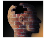
 cerebellum.

There are more connections in the cerebellum than in the whole of the rest of the brain put together, yet it is still not clear what sort of contribution it makes to thought and behaviour.

It is known that it is essential for movement, as damage to this area can produce tremor and other movement disorders – such as a condition called cerebellar ataxia.

Curiously, it also seems to be involved in almost every other form of mental activity.

If you want a reliable way of annoying anyone presenting results from a brain scanning study, put your hand up and ask what the activity in the cerebellum signifies. It almost always occurs, but is very difficult to explain with our current understanding.

The Wikipedia article is a great summary of current knowledge though, and gives an insight into an area where neuroscience is increasingly going to focus its sights as time goes on.

Link to Wikipedia article on the cerebellum.

The ‘inchoate’ science of consciousness

brick_head.jpgNeuroscientist Christof Koch manages to write an odd article on consciousness and gets an obscure word into the title of a piece published in The Scientist.

Apparently ‘inchoate’ (I had to look it up) means “partially but not fully in existence”, which pretty much sums up the article.

It starts with a brief overview of the history of consciousness and then gives a few snapshots of recent research projects, all of which seems fine until there’s a strange paragraph on a study of mice who have had their nicotine receptors altered…

While the β2 knockout animals move rapidly through a novel terrain with little exploration, animals in which nicotinic transmission has been restored in the VTA [ventral tegmental area] show more adaptive behavior that, if observed in humans, would be associated with planning and consciousness.

Quite how exploratory behaviour in laboratory mice is ‘associated’ with human consciousness eludes me right now.

As one of the few talking mice in existence, perhaps we should ask Mickey about his conscious experience and extrapolate to his smaller cousins?

Link to article ‘The Inchoate Science of Consciousness’.