Head in a vice

Scientific American has an article on migraines that takes a comprehensive look at the science of this painful and hallucinatory disorder.

The piece updates the science on migraines from the traditional but oversimplified ‘constricted blood vessels’ explanation to explore the interplay between nerves, neurotransmitters and lifestyle.

A crucial process seems to be cortical spreading depression that may be responsible, at least in part, for both the intense pain and the aura:

Aura appears to stem from cortical spreading depression—a kind of “brainstorm” anticipated as the cause of migraine in the writings of 19th-century physician Edward Lieving. Although biologist Aristides Leão first reported the phenomenon in animals in 1944, it was experimentally linked to migraine only recently. In more technical terms, cortical spreading depression is a wave of intense nerve cell activity that spreads through an unusually large swath of the cortex (the furrowed, outer layer of the brain), especially the areas that control vision. This hyperexcitable phase is followed by a wave of widespread, and relatively prolonged, neuronal inhibition. During this inhibitory phase, the neurons are in a state of “suspended animation,” during which they cannot be excited.

Neuronal activity is controlled by a carefully synchronized flow of sodium, potassium and calcium ions across the nerve cell membrane through channels and pumps. The pumps keep resting cells high in potassium and low in sodium and calcium. A neuron “fires,” releasing neurotransmitters, when the inward flow of sodium and calcium through opened channels depolarizes the membrane—that is, when the inside of the cell becomes positively charged relative to the outside. Normally, cells then briefly hyperpolarize: they become strongly negative on the inside relative to the outside by allowing potassium ions to rush out. Hyperpolarization closes the sodium and calcium channels and returns the neurons to their resting state soon after firing. But neurons can remain excessively hyperpolarized, or inhibited, for a long time following intense stimulations.

The article is remarkably comprehensive, probably as it’s written by neurologists David Dodick and John Gargus.

Link to SciAm article ‘Why Migraines Strike’ (via 3Q).

Mental illness: in with the intron crowd

Today’s Nature has an excellent feature article on the heated scientific debates over why its so hard to link genes to specific mental illnesses.

Genetics is a complex business, but psychiatric genetics even more so, because it attempts to find links between two completely different levels of description.

Genes are defined on the neurobiological level, while psychiatric diagnoses are defined on the phenomenological level – in other words, verbal descriptions of behaviour, or verbal descriptions of what it is like to have certain mental states.

There is no guarantee, and in many people’s opinion, probably no likelihood, that these ‘what it is like’ descriptions actually clearly demarcate distinct processes at the biological level.

It’s a bit like classifying people as heavy metal fans if they have five or more heavy metal albums.

By definition, there’s a biological difference between people who like heavy metal and those who don’t, but it could be a whole number of distinct differences at the level of brain function which are all just recognised as ‘being a heavy metal fan’ in day-to-day life.

Actually, psychiatric diagnosis has an additional problem, in that for some diagnoses, the same classification can be made when the people don’t share any symptoms. For example, two people could be classified as having schizophrenia / being a heavy metal fan, when they have no symptoms / albums in common.

Some psychiatric geneticists just argue that we don’t have enough data yet, because it seems that when connecting genes to psychology each gene contributes very little and the effect is when the influence of many small effect genes add up and interact.

Others argue that we should look for effects on ‘endophenotypes‘ – the cognitive building blocks of more complex mental life. So instead of trying to connect genes to a collection of ‘what it is like’ experiences, we look at how genes influence neuropsychological processes – such as the mechanisms in the prefrontal cortex that control attention.

Increasingly, some researchers are starting to suggest that the genetic results show that existing psychiatric classifications are invalid, and that we should rethink them as new data comes in.

One thing psychiatry has traditionally been very bad at though, is refining diagnoses on the basis of lab studies.

Definitions are often revised to make them statistically more reliable (i.e. so people can reliably agree what is and what isn’t a particular diagnosis), but this is not the same as having something which is a good basis for scientific enquiry.

Unfortunately, psychiatry is (ironically) a bit too emotionally attached to the traditional diagnostic categories because diagnosis is such a core part of what psychiatrists do.

Anyway, the Nature piece is an excellent guide to the debate on whether we should be attempting to link genes to the neuropsychology of mental disorder.

Link to article ‘Psychiatric genetics: The brains of the family’.

Neurowarfare and the modern Rogue Trooper

Wired has picked up on a US military report that warns of the threat posed by neuro-enhanced enemy soldiers, just released by the “Pentagon’s most prestigious scientific advisory panel”.

The full report is available online as a pdf file, and covers how pharmaceuticals and brain-computer interfaces could be used by enemies of the US to create hordes of sleep-resistant super-intelligent neurosoldiers who can kill at the speed of thought.

Obviously, I paraphrase, but it’s interesting that the report is not your usual blue-sky speculation. It actually covers the science in considerable detail.

It also discusses cultural attitudes to cognitive and brain enhancements of various sorts, and how this might affect how and why they might be used.

Non-medical applications of the advances of neuroscience research and medical technology also pose the potential for use by adversaries. In this context, we must consider the possibility that uses that we would consider unacceptable could be developed or applied either by a state-adversary, or by less-easily identified terrorist groups. In the following, we consider first the issues of what types of human performance modification might alter a military balance, and how those issues can be evaluated. We then address two broad areas where there are significant, and highly publicized, advances in human performance modification. These are the areas of brain plasticity (permanently changing the function of an individual’s brain, either by training or by pharmaceuticals), and the area of brain-computer interface (augmenting normal performance via an external device directly linked to the nervous system).

Link to Wired write-up.
pdf of report.

Brain twister

In 1941, brain specialist Russell Brain published an article about the brain in the brain science journal Brain. Owing to Brain’s extensive work on the brain, he later became editor of Brain. His work treating brain disorders and his editorship of Brain were some of the reasons he was made Baron Brain, in 1962.

Last year, Brain published a tribute to Brain’s brain article in Brain, owing to its massive impact on our understanding of the brain.

It was written by Alastair Compston.

Connected to the highways of the brain

A fantastic new study which looked at the ‘connectedness’ of the human brain has identified which aspects of the underlying network are the most important routes of communication.

The research was led by neuroscientist Patric Hagmann and combines brain imaging with network mathematics to not only visualise the brain’s network but also to understand which are the most important hubs and connections.

The study used diffusion spectrum imaging or DSI to map out the white matter wiring of the brain in five healthy individuals.

It’s a type of diffusion MRI that identifies water molecules and tracks how they move. In a glass of water, water molecules will move randomly, but when trapped inside nerve fibres, they move along the length of the fibre, allowing maps to be created from the average paths of the moving molecules.

The researchers then took the maps of fibres, as illustrated by the top image, divided the brain up into sections, and created a simplified network map, shown in the bottom image, which allowed them to mathematically test how connected the different areas were.

They used network theory, more typically used in social network analysis, which allows mathematical measures of network properties.

The researchers calculated which areas were the most connected to the rest of the network in terms of connections going directly in and out of the area, but also which areas were the most strategically important ‘hubs’.

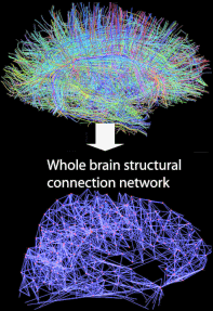
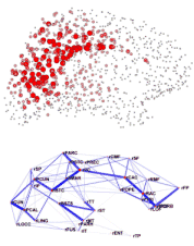
This meant the researchers could identify areas of the cortex that are the most highly connected and highly important, forming a structural core of the human brain.

You can see two of the maps on the right. The one in red illustrates which brain areas are the most highly connected. You can see it’s the area at the top and back of the brain. As you can see better on the original image, its very centrally located, like a neural mohawk.

The image in blue on the right shows the network ‘backbone’, the information highways of the brain.

What’s perhaps most interesting it that the most connected brain areas are many of those which are more active when we’re at rest, compared to when we’re engaged in a mental task that requires concentration and effort.

This has been dubbed the ‘default network’ in the scientific literature, and, rather annoyingly, the ‘daydreaming network’ by the popular press.

It’s not entirely clear what the network is for, with some studies directly linked it to ‘stimulus independent thought’ (yes, daydreaming), while others more explicitly define it as internally focused, rather than externally focused thought and cognition.

Unfortunately, most cognitive neuroscience experiments work by measuring the effect of tasks on brain function, so a brain network which seems to be switched off by any sort of task is quite hard to study. A recent study found that even the noise of the brain scanner affects it.

Link to PLoS Biology article on brain connectivity.
Link to write-up from The New York Times.
Link to write-up from Neurophilosophy.
Link to write-up from Science News.

Out of body experiences and grasping the ungraspable

This week’s ABC Radio National’s All in the Mind discusses what happens in the brain during out of body experiences, and why actions can be accurate even when our perceptions are not.

The first interview is with neurologist Olaf Blanke who discusses some of his recent compelling research, including a virtual reality experiment to induce out-of-body touch sensations in healthy participants and one with implanted brain electrodes to trigger full-blown out-of-body experiences in patients undergoing neurosurgery.

The second interview is with psychologist Melvyn Goodale, famous for his work on distinguishing the visual streams in the brain: the dorsal stream and the ventral stream.

Some of the most striking and important results from this work come from patients who have suffered damage to one or the other stream.

In the programme, Goodale talks about brain-injured patient DF, who can correctly and accurately grasp objects she cannot consciously ‘see’. The opposite has been found in other patients, who can accurately see and describe objects they cannot accurately grasp.

This suggests that these two visual pathways, although complimentary, are specialised for different things, one for identifying objects, and the other for working out where they are and how to manipulate them.

The different function of the two pathways can also be demonstrated in healthy people as well.

You may recognise the visual illusion on the left, sometimes called the Titchener or Ebbinghaus illusion. The two circles in the middle are actually the same size, but look different due to their context.

Researchers have created a graspable version of the illusion by putting hoops on a flat surface.

When they’ve measured how people adjust their fingers to pick up the middle circles, they find that we don’t over or underestimate the size. Our fingers are always perfectly adjusted to the actual size.

In other words, it seems that while our perception is fooled by the illusion, our actions aren’t, showing how the specialisation of each visual stream can be seen in everyone.

There’s now a minor cottage industry of research attempting to understand exactly what influences the effect.

UPDATE: “All in the Mind has been honoured with the Grand Award at 2008 New York Radio festivals for best entry across all categories, as well as a Gold World Medal in the Health / Medical category”. – I’m sure it won’t come as a surprise to most Mind Hacks readers but fantastic to have it recognised by the non-initiated!

Link to AITM on out-of-body experiences and other tricks of consciousness.

Back from the dead

A scene from a thousand horror movies, retold in the medical literature, with an additional lesson about the correct use of cerebral perfusion and angiography in diagnosing the brain dead patient.

Presumably, learnt shortly after the doctors had stopped screaming.

I love the use of the phrase “the situation became confusing”, just after the dead guy starts moving again.

Unusual movements, “spontaneous” breathing, and unclear cerebral vessels sonography in a brain-dead patient: a case report.

Bohatyrewicz R, Walecka A, Bohatyrewicz A, Zukowski M, Kepiński S, Marzec-Lewenstein E, Sawicki M, Kordowski J.

Transplant Proc. 2007 Nov;39(9):2707-8.

A patient with a brain injury fulfilled all clinical criteria for brainstem death diagnosis. Two standard sets of tests were performed; according to Polish regulations, the patient could be declared brain dead. However, shortly after the completion of the tests and before the final brain death declaration, 6 triggered “assisted” breaths/min were noticed. After careful analysis of the ventilator settings, it was concluded that low trigger sensitivity and airway pressure oscillations during heart contractions were the reasons.

Additionally, a few minutes later, spontaneous jerking movements of lower limbs and clonic movements of neck muscles secondary to painful stimuli were noticed. The situation became confusing; therefore, cerebral Doppler sonography was performed, showing circulatory arrest in both of the internal carotid, middle cerebral, and left vertebral arteries. The basilar artery was not visualized. Forward flow with increased pulsatility was recorded in extracranial and intracranial segments of the right vertebral artery. Cerebral circulatory arrest was still uncertain; therefore, the diagnostic procedures were completed with conventional cerebral angiography, which showed a lack of cerebral blood flow.

Finally, the patient was declared brain dead; kidneys and bones were harvested. Cardiogenic oscillations associated with incorrect low ventilator trigger settings may falsely suggest persistence of breathing efforts in a brain-dead patient. In the case of any unusual events during brain death diagnosis, cerebral perfusion tests should be performed with cerebral angiography as the “gold standard.”

Link to PubMed entry.

The fMRI smackdown cometh

Over the last few months, the soul searching over the shortcomings of fMRI brain scanning has escaped the backrooms of imaging labs and has hit the mainstream.

Numerous articles in hard hitting publications have questioned some common assumptions behind the technology, suggesting a backlash against the bright lights of brain scanning is in full swing.

There are two strands to this debate, and both stem from the fact that the technology and conceptual issues of brain imaging are incredibly complex.

To fully understand what happens during a brain imaging experiment you need to be able to grasp quantum physics at one end, to philosophy of mind at the other, while travelling through a sea of statistics, neurophysiology and psychology. Needless to say, very few, if any scientists can do this on their own.

So the first strand involves how brain imaging experiments are reported in the media. Under the sheer weight of conceptual strain, journalists panic, and do this: “Brain’s adventure centre located”.

It’s a story published this morning on the BBC News website based on an interesting fMRI study looking at brain activity associated with participants choosing a novel option in a simple gambling task. But out of the four words of the headline, only the first is accurate.

And this leads to the second strand of the debate, which, until recently, has been largely conducted away from the media’s gaze, amongst the people doing cognitive science themselves.

It starts with this simple question: what is fMRI measuring?

When we talk about imaging experiments, we usually say it measures ‘brain activity’, but you may be surprised to know that no-one’s really sure what this actually means.

Neuroscientist Nikos Logothetis published an important paper in Nature a couple of weeks ago explaining exactly what we know so far about the link between what brain scans measure and what the brain is actually doing.

It’s very wide-ranging and includes lots of grit-your-teeth hardcore neurophysiology, but is, I think, essential reading if you’re neuroscientifically inclined.

It focuses on BOLD, the signal that reflects the ratio of oxygenated and deoxygenated blood measured by fMRI, and the fact that it can be altered by a huge range of different biological process and neural firing patterns.

One of the main points of the paper is that the brain is not simply an array of tiny localised processors, but it is more like an an ecosystem of communication.

Activity can result from sending more signals, trying to send less, or, from what seems to be particularly important – maintaining a balance of excitation and inhibition.

Furthermore, it seems that a great deal of neural activity is not from neurons that might be directly involved in a task, but from ‘neuromodulation’ – general processes of management and coordination, often linked to attention. This can wax and wane, can spread like ripples and can occur in all sorts of non-linear ways that makes interpretation difficult.

What this means is that brain imaging experiments need to be carefully designed to control for these effects, but this entirely depends on our understanding of the effects themselves.

In other words, our understanding of what brain scanning data tells us evolves over time. A study conducted ten years ago might mean something different now.

An article in Science, published in the same week as Logothetis’ paper, reports on new statistical methods for interpreting imaging data, a different issue again.

The latest edition of The New Atlantis has an article that attempts to come to grips with some of the philosophical aspects of brain imaging experiments, in terms of the conceptual limits in inferring mental states from biological changes.

I have to say, it’s a bit miscued in places, assuming that brain imaging relies on ideas about brain modularity (which it doesn’t) and seemingly confusing it with the notion of pure insertion, and suggesting some rather strange notions about mental causation, but it has many good points and is worth a read.

It’s important that these sorts of issues come to light, because it hopefully heralds a time of increased caution in our interpretation of brain scans – and that goes for scientists, the media and the general public.

This is essential, because this data is starting to be used, literally, in life or death decisions.

The same issue of The New Atlantis has an article on neuroimaging that discusses the ethical dilemmas in applying this imperfect technology to legal decisions concerning capital punishment.

Link to Logothetis on ‘What we can do and what we cannot do with fMRI’.
Link to Science article ‘Growing pains for fMRI’.
Link to New Atlantis on ‘The Limits of Neuro-Talk’.
Link to New Atlantis on Neuroimaging and Capital Punishment.

Gazzaniga on split-brains and bioethics

Michael Gazzaniga, one of the founding fathers of cognitive neuroscience and a pioneer of ‘split brain’ research, is interviewed on this week’s ABC All in the Mind where he talks about the use and abuse of ‘left brain – right brain’ metaphors and how our understanding of free will is impacting on the law.

Gazzaniga was a student of Roger Sperry, who won a Nobel prize for his work on ‘split-brain patients’, people who had the two cortical hemispheres of the brain functional separated by neurosurgery to cut the corpus callosum in an attempt to treat otherwise untreatable epilepsy.

One of the amazing things was that while the people didn’t feel any different, it was easy to demonstrate that the each hemisphere processed things in quite different ways and each was, to a certain extent, independently conscious.

The interview discusses some of this early research, and asks how much of the popular ‘left brain – right brain’ rhetoric that gets thrown around actually stands up to scientific scrutiny. I think you can guess, but it’s good hearing it from the man himself.

Gazzaniga also talks about one of his other interests – neuroethics, and particularly the effect that a neuroscientific understanding of free will is having on our concepts of legal responsibility.

I was interested to read that US judges can now take courses in neuroscience to help them makes sense of the sometimes counter-intuitive findings in cognitive science.

As it happens, Gazzaniga’s new book Human: The Science Behind What Makes Us Unique is published today. If you want a taster an Edge article by Gazzaniga from a few months ago seems to be taken from it.

The AITM Blog also has some bonus audio of Gazzaniga discussing his experience of being on George Bush’s bioethics council when the President was vetoing stem cell cloning.

Link to AITM interview with Michael Gazzaniga.

Synapse structure varies across species

The New York Times covers new research which has found significant cross-species variation in the structure of the synapse – the chemical ‘connection points’ that allow neurons to communicate.

The study itself has been published in Nature Neuroscience and the full text is available online for those who want the in-depth science.

A whole new dimension of evolutionary complexity has now emerged from a cross-species study led by Dr. Seth Grant at the Sanger Institute in England.

Dr. Grant looked at the interconnections between neurons, known as synapses, which until now have been regarded as a standard feature of neurons.

But in fact the synapses get considerably more complex going up the evolutionary scale, Dr. Grant and colleagues reported online Sunday in Nature Neuroscience. In worms and flies, the synapses mediate simple forms of learning, but in higher animals they are built from a much richer array of protein components and conduct complex learning and pattern recognition, Dr. Grant said.

The finding may open a new window into how the brain operates. “One of the biggest questions in neuroscience is to answer what are the design principles by which the human brain is constructed, and this is one of those principles,” Dr. Grant said.

The paper itself doesn’t mention the issue, but I wander what implications this might have for the generalisation of animal experiments to humans.

The majority of cellular-level neuroscience research is done on animal tissue. While some of this focuses on the molecular level, where differences in the structure of, let’s say, ion channels, would be easily apparent in comparison to humans, some studies simply look at the ‘synapse’ as the smallest functional unit.

In fact, a considerable amount of neuroscience research is done on the 1mm long microscopic worm C. elegans and the fruit fly, drosophila. This new research suggests that neuroscientists may need to be additionally cautious when assuming that the findings relate to general laws that might apply in humans.

UPDATE: Neurophilosophy has a great write-up of this study, which discusses it in more detail.

Link to NYT article ‘Brainpower May Lie in Complexity of Synapses’.
Link to PubMed entry for scientific paper.
Link to full text.

Wake up and smell the caffeine

New York Magazine has a wonderful article on the culture, controversies and pharmacology of caffeine – the world’s most popular psychoactive drug.

Ranging from the recent upturn in coffee’s popularity and its inevitable effect on our caffeine consumption to the science of its neurological effects, the article manages to capture some of the key debates about the tremor inducing buzz substance.

One particularly interesting part touches on research that suggests that, like the effect of nicotine, the lift for regular users may be nothing more than withdrawal symptoms being soothed to bring us back to baseline.

That all said, what if the uptick in energy, alertness, and smarts we feel after drinking a cup of coffee isn‚Äôt a real uptick at all? What if it‚Äôs an illusion? A group of cutting-edge caffeine researchers believes that might be the case…

When Griffiths and Juliano teamed up to review 170 years of caffeine research, much of which confirmed the drug’s reputation as a brain booster, they noticed a pattern: Most studies had been done on caffeine users who, in the interest of scientific rigor, were deprived of the stimulant overnight. Because caffeine withdrawal can commence in just twelve hours, by the time each study’s jonesing test subjects were given either caffeine or a placebo, they had begun to suffer headaches and fatigue.

For the half that received the stimulant—poof!—their withdrawal symptoms vanished. The other half remained uncaffeinated, crabby, and logy, and guess which group scored higher on cognitive tests time after time? The boost the test subjects who got the caffeine felt may have simply been a function of having been deprived of the drug.

Link to ‘The Coffee Junkie‚Äôs Guide to Caffeine Addiction’.

Fantastic introduction to MRI brain scanning physics

Magnetic resonance imaging is the most popular method for scanning the brain both for research and for clinical investigations. I’ve just found a wonderfully written article that gives a great introduction to the physics of how MRI scanners work.

It is both clearly written for the non-specialist and fantastically complete. This is a rare and valuable combination.

There are some other guides to MRI physics which are also wonderfully written but most lack the sufficient detail that would bring you up to ‘entry level’ in the field.

For example, How Stuff Work’s guide to MRI is a great place to start, but it won’t tell you about why and how T1 and T2 imaging are different, or any of the other things you need to know to understand the fundamentals of MRI technology.

You don’t need to know maths to understand the article (the downfall of most ‘introductory’ guides to MRI) and the author uses wonderfully clear analogies throughout.

The article is written by radiologist Robert Pooley, who should give himself a pat on the back for such a great job. It was published as an open-access paper in the journal RadioGraphics. Perfection!

Link to article ‘Fundamental Physics of MR Imaging’.

Hash high in cannabidiol but varies widely

In light of research showing that an ingredient in cannabis, cannabidiol, seems to actually reduce the risk of psychosis, I speculated previously on Mind Hacks whether smokers might be attracted to high-cannabidiol dope.

A study of UK street cannabis published in the Journal of Forensic Sciences suggests that cannabis resin (hashish) has the average highest rates of cannabidiol, while ‘skunk’ and imported herbal cannabis (weed) have the lowest.

For people who take cannabis, it’s not the cannabidiol that makes you ‘high’, it’s mainly a substance called tetrahydrocannabinol or THC.

There’s accumulating evidence that THC increases the risk of psychosis, while cannabidiol reduces it – so the ratio of the two substances in the street drug might give a ‘risk profile’ in terms of mental health.

‘Might’ is the operative word here, as the research is still preliminary and the studies are still largely correlational with regard to cannabidiol-to-THC ratio and psychosis-like symptoms.

However, if this does turn out to be case, the new survey of UK street cannabis suggests that, on average, cannabis resin has higher levels of cannabidiol, with the implication that this might be less risky in terms of developing schizophrenia or other psychotic disorders.

This finding is an average over all the samples, however, and the study also found that resin had quite a bit of variability with regards to cannabidiol-to-THC ratio.

However, imported herbal cannabis and skunk was generally very low in cannabidiol. Additionally, skunk also had about 6 times the THC content of normal weed, making it especially potent.

The study concludes:

This study suggests that cannabis in England in 2005 remains a very variable drug with unpredictable pharmacological and psychological activity. The potency (THC content) of the cannabis varies widely, as does the content of other cannabinoids, especially in herbal cannabis and cannabis resin. The average potency within the country appears to be increasing, but large variations remain within and between different areas of the country.

CBD affects the pharmacological qualities of THC and reduces it psychoactive potential. The relative proportions of THC and CBD in resin are wide ranging, supporting the view that the potential effects of resin cannot be judged by measuring the THC content alone. The resin samples were all similar in appearance and gave the user no indication of their cannabinoid content.

Of the three principle forms of cannabis, sinsemilla [skunk] commonly had the highest THC content and almost totally lacked CBD. Had CBD been present it would have reduced the psychoactive potential of this material. In addition to having increased in potency, sinsemilla also appears to have become the most widely used form of cannabis. The current trends in cannabis use suggest that those susceptible to the harmful psychological effects associated with THC are at ever greater risk. This is due to the combined rise in potency and popularity of sinsemilla and the absence of CBD in this product.

The lead scientist in the study is called Professor Potter. Do with that fact as you will.

Link to abstract of Journal of Forensic Sciences study.

Like a bullet in the head

Neurophilosophy has collected some of the most unusual cases of penetrating brain injury from the medical literature, with x-rays that illustrate how some of the most curious objects can end up on the wrong side of the bony brain protector.

You may recognise a couple that we’ve noted before on Mind Hacks, but this is a far more complete and frankly quite surprising collection.

The most amazing is the case of a “32-year-old Caucasian male with a history of repeated self-injury drilled a hole in his skull using a power tool and subsequently introduced intracerebrally a binding wire from a sketchpad”.

A striking, and, in some places, stomach churning collection of case studies.

Link to Neurophilosophy on unusual penetrating brain injuries.

The brains of dead Russian geniuses

What makes a man a genius? Russian neuroscientists were pondering this exactly this question in the early 1900s and did exactly what seemed sensible at the time – they collected and dissected the brains of some of the greatest cultural figures in a huge collection called ‘The Pantheon of Brains’.

It’s a fascinating story told in a recent article published in the medical journal Brain. Amazingly, the last brain was only added in 1989.

Rather fittingly, the collection contains the brains of some of the Russia’s greatest psychologists and neuroscientists and has many curious aspects to it, such as the mysterious death of its founder. After death, his brain was immediately added to the collection.

In 1927, Bekhterev came up with a plan to organize ‘The Pantheon of Brains’ in Leningrad in order to collect elite brains. It was a severe irony of fate that precisely when the question about creating the Pantheon had been positively solved, the very initiator of this creation, Bekhterev, suddenly passed away. The circumstances are still questionable.

On December 17, 1927, the First All-Union Congress of Neuropathologists and Psychiatrists was held in Moscow. Bekhterev, along with L. S. Minor and G. I. Rossolimo, was elected as honourable chairmen of the congress. On December 23rd, the last day of the congress, Bekhterev gave a presentation during the afternoon session. In the evening, symptoms of a gastrointestinal disorder started and 24 hs later, Bekhterev died of (as officially stated) acute heart failure. Without any further post-mortem pathoanatomical investigation, his brain was removed, in accordance with his will, and his body was cremated the next day. However, the idea did not fade away.

In 1928, the neuroanatomical laboratory of Vogt and his Russian colleagues were reorganized into the Moscow Brain Research Institute, where the structured collecting and mapping of the brains of famous Russians started. Bekhterev did not see his plan come to fruition, but his own brain enriched the collection of the Moscow Institute (the weight of his brain was 1720g). The collection acquired the brains of Soviet politicians, famous writers, poets, musicians, etc.

It is not surprising that these included the brains of prominent Russian neuroscientists, such as neurologist, G.I. Rossolimo (1860‚Äì1928) – 1543g; physiologist, I.P. Pavlov (1849‚Äì1936) – 1517g; neurologist, M. B. Kroll (1879‚Äì1939) – 1520g; psychiatrist, P. B. Gannushkin (1875‚Äì1933) – 1495g; psychologist, L.S. Vygotsky (1896‚Äì1934). During the Soviet period, the work of the Moscow Brain Research Institute continued behind closed doors.

The collection was still expanding as recently as 1989, when it acquired the brain of A.D. Sakharov [A. D. Sakharov (1921–89) was an eminent Soviet nuclear physicist, dissident and human rights activist. He was an advocate of civil liberties and reforms in the Soviet Union. He was awarded the Nobel Peace Prize in 1975] — 1440g.

You gotta love the fact that the authors have added exactly how much each person’s brain weighed.

Sadly, the full text isn’t available online, although Brain does fully release articles after a set amount of time (a year I think) so it should eventually see the light.

Link to PubMed entry for article.

Ecstasy’s impact

I’ve just noticed this review article that concisely reviews what we know about how the street drug ecstasy (MDMA) affects the function of the brain.

In terms of life-threatening physical damage, MDMA is a great deal safer than most other recreational drugs including alcohol and tobacco, but there is increasing evidence that it impacts on memory, and the effect seems to be related to dose.

In other words, the more ecstasy you take, the more likely memory problems will be worse.

The neuropsychology of ecstasy (MDMA) use: a quantitative review.

Hum Psychopharmacol. 2007 Oct;22(7):427-35.

Zakzanis KK, Campbell Z, Jovanovski D.

A growing number of empirical studies have found varying neuropsychological impairments associated with use of 3,4-methylenedioxymethamphetamine (MDMA) use. We set out to determine to what extent neuropsychological abilities are impaired in MDMA users. To do so, meta-analytical methods were used to determine the magnitude of neuropsychological impairment in MDMA users across pre-specified cognitive domains. We found that cognitive impairment secondary to recreational drug use may result in what might be described as small-to-medium effects across all cognitive domains with learning and memory being most impaired. We also found that total lifetime ingestion of MDMA appears to be negatively associated with performance on tasks ranging from attention and concentration to learning and memory. Implications and limitations of these findings are discussed.

Sadly, the full-text of the paper isn’t freely available online, but the main punchlines are in the summary.

Link to PubMed entry for paper.